What is a 3-Tesla MRI?
Magnetic resonance imaging, or MRI for short, provides the images of the body's internal structures by using radio waves and magnets. Medical professionals can use an MRI to examine what's going on inside a person's body without having to perform an invasive procedure. MRI, unlike X-rays, employs non-ionising radiation.
A Tesla is a unit of measurement for magnetic field strength. 3-Tesla MRI, or 3T MRI, employs extremely powerful magnets to generate a magnetic field of 3 Tesla. The strength of this magnetic field is twice of that of the fields used in traditional MRI scanners. Therefore, the image obtained using 3T MRI is clearer and more complete than that acquired through traditional MRI. In addition, stronger magnetic fields generate better pictures of organs and soft tissues than standard MRI scanners. Additionally, the 3-T MRIs are fast and comfortable (spacious).
How to prepare for a 3T MRI scan?
There is no specific preparation for an MRI, unless specified by the doctor.In general,
- Carry your medical history as well as any previous scans with you. This information will assist the radiologist in planning and generating an appropriate report.
- Remove all metallic objects, including jewellery, belts, watches, keys, and wallets. Also, some of the cosmetics may have metal traces, so it is best to remove your make-up prior to the scan.
- Prior to the MRI, you will be required to fill out a screening form in which you will be asked about anything that may pose a potential health risk or interfere with MRI imaging (e.g., a metal implant, pacemaker, foreign metal objects, or bullet wounds).
- If you think you are claustrophobic, please notify the radiologist.
- Female patients should inform their doctor and radiologist about their pregnancy or if they intend to become pregnant.
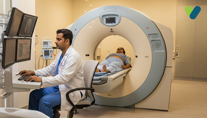
What to expect during the 3T MRI scanning?
- You must lie down on your stomach or your side on the scanner table that slides through the scanner. The technician will ensure that you are positioned correctly.
- Once you are in position, the technician will begin the scan. Hearing some noises during the procedure is absolutely normal.
- You must lie down still for a successful MRI scan. If you experience discomfort, you can communicate with the radiographer through the scanner's embedded intercom.
- Sometimes a contrast dye can be given orally or by injection before the scan to get clearer images of the hard-to-access parts of the body's organs and their nearby areas. Since the dye takes about 30 minutes to get absorbed in the system, you may have to rest and limit your movement after taking it.
- The radiographer will check the captured images after scanning, but if they determine that additional images are required, you may be asked to retake the test.
What are the risks associated with the 3 T MRI scan?
While MRI scans are usually thought to be safe, there are some risks involved with 3T MRI scanning, which include:- Magnetic interference: Metal objects on or within the body can be pulled or shifted by the powerful magnetic field generated by a 3T MRI, causing injury or damage.
- Claustrophobia: During the scan, some individuals might feel anxious or claustrophobic, particularly if they are positioned inside a narrow tube.
- Pregnancy: If you are currently pregnant or suspect that you may be pregnant, you should let your doctor or the MRI technician at the MRI centre know prior to the imaging procedure since the magnetic field may interfere with the foetus's growth.
- Contrast material: There is a slight possibility of an allergic reaction or kidney damage if contrast medium is used during the scan.