Brain PET is a positron emission tomography technique used to examine the distribution of exogenous radiolabelled chemical agents and brain metabolism. PET is employed to analyse the chemical emissions that have been put into the bloodstream and are radioactively marked. Computer processing of the brain PET emission data results in multi-dimensional images of the distributed substances in the brain.
Explaining Brain PET scan
The imaging test called a brain positron emission tomography (PET) scan helps medical professionals to deduce the health of your brain. Following the bloodstream absorption of radioactive tracer chemicals, the scan records brain's activity. The tracers are connected to the chemicals, such as glucose. Glucose serves as the brain's principal fuel source. Active brain regions utilise glucose more quickly than inactive ones. In most cases, it is an outpatient operation. This means when you are done with the test procedure, you can get back to your daily routine.Purpose of a Brain PET scan
The examination gives exact details regarding the size, composition, and brain operations. In contrast to standard scans, a brain PET scan provides doctors with a picture of your brain's structure and functionality.This enables the doctors to:
- Examine for cancerous and non-cancerous tumours.
- Evaluate spread of cancer in other parts of the brain, identify dementias like Alzheimer's disease, and distinguish it from other disorders.
- Prepare for the operation for epilepsy.
Preparation before the Brain PET scan
You will receive detailed instructions from your doctor on how to get ready for a brain PET scan. You must share the details about all the medicines you are taking, the prescription drugs and even the supplements, with your doctor. Before the treatment starts, you will be asked to fast for 8 hours. However, you can drink water like usual. Inform your doctor if you think you might be pregnant or if you are already pregnant. Your foetus could be in danger from the exam.Any medical issues you already have should be disclosed to your doctor. For instance, particular instructions for the test will probably be given to individuals with diabetes. Their level of blood sugar can be negatively impacted by fasting prior to the test. You need to wear a hospital gown before the test and take off all of your jewellery and metal accessories.
How is a Brain PET Scan Performed?
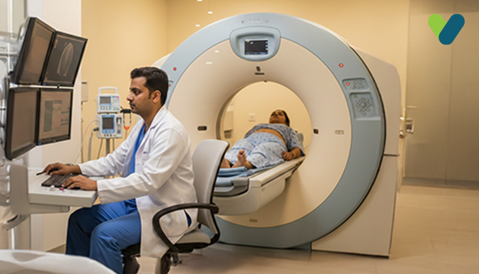
A technician guides you into the treatment room and places an intravenous catheter (IV) into your hand. A special dye containing radioactive tracers will be administered during this process. The scan won't begin right away because your body requires some time to absorb tracers as your blood flows through your brain. This scan normally takes an hour. You will then get the scan done under the supervision of a technician. To do this, you must lay down on a PET machine table and position yourself appropriately. To finish the scan, the table enters the device smoothly.Remain still while you are going through the scan. The professional will ask you to restrict your movement at certain intervals. The activities of the brain are captured live by the scans. They can be captured in still or moving photographs. In regions with higher blood flow, the accumulated tracers can be seen.
You will be taken away from the machine once the desired photograph is taken there, ending the scanning procedure.
Observations after the Brain PET Scan
Drinking enough water following the test will help your body in removing the tracers. Within two days, the majority of tracers will leave your body. Apart from that, you can carry on with your life as normal unless your physician instructs you otherwise.An expert with experience in reading various PET scans will deduce the readings of the images while sharing the results with your specialist. During a subsequent appointment, your specialist will discuss the findings.
Risk factors for the Brain PET Scan
Although radioactive tracers are used in the scan, the dose is quite low. It isn't high enough to interfere with the body's natural functions.The test's hazards are negligible in light of the potential benefits of the findings. Pregnant women, or women suspecting to be pregnant, or those who are still breast feeding, however, should not have any kind of PET CT scan brain because radiation is thought to be harmful to foetuses and babies.
Additional dangers include uneasy emotions, especially if you have claustrophobia or needle anxiety.
Benefits of the Brain PET Scan
Some of the benefits related to brain PET scans are discussed below.Early detection of diseases: PET CT brain scan can examine minor changes in the function of the brain, which may be the cause of neurological disorders such as Alzheimer's disease, Parkinson's disease, and various brain diseases. Early detection can help doctors to start treatments before the disease progresses.
Accurate diagnosis: Brain PET scans generate the images of the brain in high resolution, which assists doctors in accurately detecting and differentiating between various brain diseases.
Treatments: Brain PET scans can assist doctors in determining the appropriate treatment plan for each patient based on the specific areas of the brain that are affected.
Monitoring treatment effectiveness: Brain PET scans can help doctors monitor the effectiveness of treatment for neurological conditions and adjust treatment plans accordingly.
Interpreting the results of the Brain PET scan
You can observe a variety of colours, ranging from deep red to dark blue, in the images produced by brain PET scans. Warmer hues like yellow and red, which represent active brain regions, appear on the human eye. These scans will be examined by your doctor, who will look for any anomalies.For instance, a brain tumour will appear on the PET scan as a dark patch. The areas of the brain that are larger than normal will show darker on the scan if the person has Alzheimer's disease or other types of dementia. The dark regions in both of these images denote brain regions that are damaged.
Your physician will discuss the results of your individual scan with you to clarify all the problems.