What is a CBCT scan?
Cone-beam computed tomography, also known as a CBCT scan, is a type of imaging technology used mostly by dental practitioners. Unlike traditional computed tomography (CT) systems, CBCT revolves around the patient, gathering information (multiple images) through a cone-shaped X-ray beam from various angles. The information collected is then processed to produce a three-dimensional image of the patient's dental structure, oral and maxillofacial region, that is, jaw, mouth, and neck, as well as the ears, nose, and throat (ENT).This imaging technology is commonly used by dental professionals to obtain a comprehensive view of the patient's anatomy. Therefore, it is often called a dental CBCT scan.
Why is the CBCT test performed?
Radiologists and dentists are increasingly using CBCT for a variety of clinical applications, including:Dental imaging
CBCT is used to evaluate the exact position of jaw pathologies, including inflammatory lesions and tumours, and the actual location of the impacted tooth, prior to oral or maxillofacial surgery.CBCT can also be used in implant dentistry, forensic dentistry, temporomandibular joint imaging, endodontics (e.g., root canal), periodontics (e.g., gum treatments), and orthodontics (e.g., teeth braces). Other uses of CBCT include:
Breast imaging
To overcome the limitations of two-dimensional mammography, cone-beam CT technology is used for breast imaging. It gives a 3-dimensional view of the breast tissues, making it easier to distinguish between normal and pathological tissues.Lung imaging
CBCT technology is used to guide transbronchial biopsy of peripheral pulmonary lesions and to ablate target lesions during bronchoscopy. This imaging technique aids in the guidance and accuracy of the procedure.Liver imaging
CBCT is used to assess the effectiveness of transcatheter arterial chemoembolization in the treatment of hepatocellular carcinoma. This imaging technique aids in determining the amount of drug retained within the tumour.What to prepare for a CBCT scan?
There is no need to prepare for a CBCT examination. Before the examination begins, you may be requested to take out any metallic objects that could interrupt the imaging, including jewellery, hairpins, eyeglasses, and hearing aids. Though the removable dental work might have to be removed, bringing those to the examination is recommended because your dentist might need to examine them as well.If a woman is pregnant, she should always notify her dentist or oral surgeon.
How is the CBCT scan carried out?
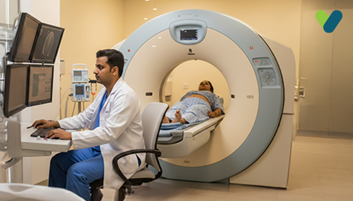
Based on the type of CBCT scanner used, you may be required to remain seated in the exam chair or lay on the exam table. Your dentist will position you in order to centre the beam on the target region. You will be asked to remain as still as possible while the X-ray source and detector rotate around you.This can take anywhere from 20 to 40 seconds for a full mouth x-ray, which images the complete mouth and dental structures, and maybe less than 10 seconds for a regional scan, which concentrates on a particular area of the upper or lower jaw.
What are the risks associated with a CBCT scan?
There is, at all times, a slight possibility of developing cancer from excessive radiation exposure. The advantage of an accurate diagnosis, on the other hand, far exceeds the risk of CT scanning.Since children are much more vulnerable to radiation exposure than adults; they should only have a CT scan if it is mandatory for a diagnosis. They shouldn't undergo repeated CT scans unless absolutely necessary. CT scans for children must always be performed using a low-dose technique.

