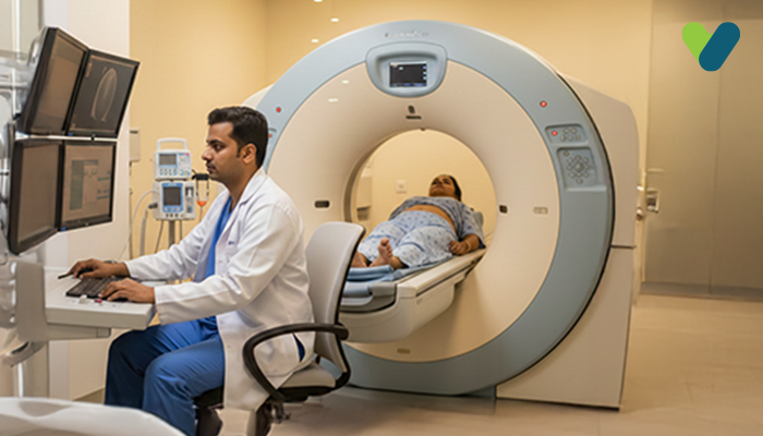
What exactly is a CT scan of the chest?
A chest CT scan is an imaging procedure that creates detailed images of the structures and organs within your chest using X-ray and computer technology. These scans provide more detailed images than standard X-rays, allowing for a more accurate diagnosis of chest organ diseases and injuries.During a CT scan, an X-ray beam moves in a circular pattern around your body, taking several images or "slices" of the lungs and interior of the chest. These images are then processed by a computer and displayed on a monitor. In some cases, a contrast dye may be given during the test to help certain areas of your body stand out more in the image.
Why is a chest CT scan needed?
A chest CT scan may be performed for the following reasons:- Examine any abnormalities discovered on chest X-rays.
- Assist in the diagnosis of the chest diseases symptoms such as coughing, chest pain, or shortness of breath.
- Check to see if tumours are getting better with therapy.
- Assist in the planning of radiation therapy.
- Detect and assess the extent of tumours that arise in the chest or tumours that are spread from elsewhere in the body.
- Evaluate for chest injuries, blockages, infections, and intrathoracic bleeding.
- Tumours, both benign and malignant
- Tuberculosis
- Pneumonia
- Chronic and interstitial lung disease
- Cystic fibrosis
- Congenital abnormalities
- Bronchiectasis
- Inflammation or other pleural diseases
How is a chest CT test performed?
The test is carried out as follows:- You will be required to change into a hospital gown.
- Next, you will be asked to lie down on a table that slides through the scanner. Once you are inside the machine, the X-rays will rotate around you to take multiple pictures of the chest area.
- You will be instructed to remain still inside the scanner to avoid any blurring of the images. You may also be asked to hold your breath for a brief period of time.
How to get ready for the chest CT scan?
- You will be required to remove all metallic objects, including jewellery, piercings (if any), dentures, and eyeglasses.
- Some people are allergic to contrast material and might have to take medication before their test to receive it safely.
- If contrast is being used, you may be instructed not to consume anything for about 4 to 6 hours prior to the test.
What are the risks associated with a CT scan of the chest?
CT scans are controlled and monitored to ensure that the least amount of radiation is used. CT scans use low concentrations of ionising radiation, which may cause cancer as well as other defects; however, the risk posed by any single scan is minimal. The risk grows as more studies are conducted.Iodine is the most commonly used type of contrast material. If an individual having an iodine allergy receives this type of contrast, they may experience sneezing, nausea, vomiting, itchiness, or hives. In rare instances, the dye can end up causing a potentially fatal allergic reaction known as anaphylaxis. If you have any breathing difficulties during the test, please inform the scanner operator right away.
The dye may be harmful to the kidneys in people who have kidney problems. Special precautions may be taken in these cases to ensure the contrast is safe to use.

