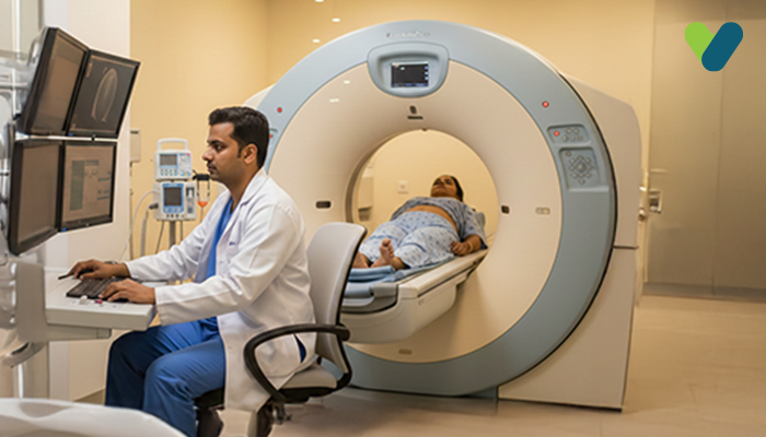
What is a DEXA scan?
You can evaluate your bone mineral content and decay with a DEXA scan, a more precise form of X-ray imaging. There is a higher risk of fractures in the bones and osteoporosis if the density of your bones has declined.DEXA stans for dual energy X-rat absorptiometry. It is also referred to as bone densitometry. This method’s commercial method first began in 1987.
How does DEXA scan work?
To reach the target bones, it fires two beams of X-rays with various peak energies. Soft tissue absorbs the first peak, while bone absorbs the second. Your bone mineral content is calculated by deducting the amount of fluid absorbed by soft tissues from the total amount absorbed. Compared to a standard X-ray, the exam is quicker, less intrusive, and more accurate. Very little radiation is used in this process. The best method for determining postmenopausal women's mineral content of bones, according to the World Health Organization (WHO), is DEXA.Uses of DEXA test
When determining who could need a DEXA test, healthcare professionals consider several different criteria. If you are older than fifty, have experienced a fractured bone, or suffer from other disorders that cause risks to your bone health, medical professionals frequently advise getting a DEXA test to evaluate your risk of osteoporosis and fractures.Women begin to lose bone mass sooner and more rapidly than men. Therefore, healthcare professionals typically advise women to obtain a DEXA scan to check for osteoporosis at an earlier age than men.
If you possess any of the following risk factors for fractures or osteoporosis, your physician might advise a DEXA scan. They include:
Increased age – As people age, they typically lose bone density. Starting around 65 years (for women) and 70 years (for males), the National Osteoporosis Foundation advises those at average risk of fracture or osteoporosis to undergo a DEXA scan.
Previous fracture injuries – The occurrence of a broken bone, particularly after the age of 50, may indicate a higher risk of osteoporosis. Bones with less thick pores are more brittle. Family history – You may be more susceptible to bone deterioration if you or a family member has experienced osteoporosis or multiple fractures.
Medicines – Some medicines, including cancer treatments, drugs used after organ transplants, and steroids such as prednisone, may degrade your bones. General health – Your risk of fracturing a bone can increase if you have several chronic medical conditions. Lupus, Rheumatoid arthritis, liver illness, diabetes, and kidney disease are examples of chronic disorders.
A DEXA scan may also be requested by medical professionals to evaluate bone density in conditions including;
• Changes in the condition of your bones (bone health). • Keep an eye on how you are responding to any treatment, for instance, a drug for osteoporosis. • Examine your body's composition, including its proportions of muscle and fat. • Have a high rate of bone turnover, which is seen by the presence of too much collagen in specimens of urine. • Possess a thyroid disorder including hyperthyroidism. • Possess a parathyroid disorder including hyperparathyroidism. • Have suffered a fracture following only minor trauma. • Have experienced vertebral fractures or other osteoporosis-related symptoms, as shown by x-rays. • History of hip fracture (personal or maternal).
Preparation required for the DEXA test
Prior to undergoing a DEXA test, the majority of individuals can continue with their regular schedule. Unless your provider instructs you otherwise, consume food, liquids, and any prescribed medications as usual. You will be required to answer questions on an application about your current health, any medicines you are currently taking, your past or current smoking record, and the family's record of fractures.Common procedures before the test include:
- The day before your DEXA test, discontinue calcium tablets. This consists of multivitamins as well as antacids that are frequently used to minimise heartburn.
- Put on loose garments. Choose clothes without metallic zippers, buckles or buttons if possible. A comfortable top and trousers would be the right choice.
- You could be asked by the technician to take off any jewellery or other objects, such as the keys, that might contain metallic materials. For the examination, you could be provided with a medical gown.
- Because DEXA tests emit low quantities of radiation, let your doctor know if you are pregnant. To safeguard the growing foetus, medical professionals suggest avoiding all exposure to radiation throughout pregnancy.
- If you've undergone computed tomography (CT) scan that required the administration of a contrast agent or a barium exam, inform your physician in beforehand. In such cases before setting up a DEXA scan, they could ask patients to postpone it for several days.
Expectations after a DEXA test
This is a rapid and painless procedure. After the DEXA test, you can continue with your regular tasks right away. The data will be reviewed and a report will be written by experts qualified to analyse DEXA scans will be given to your healthcare physician. Your doctor will go through your test results with you and aid you in understanding how they affect your health. You can decide how to maintain the strength of your bones with the assistance of your healthcare expert. Additionally, they might suggest lifestyle and dietary modifications that could help minimise the risk of fracture.Benefits of DEXA test
- A DEXA scan is a rapid, painless, and easy procedure.
- Anaesthesia is not necessary.
- Radiation is used in quite small amounts.
- The current gold standard for diagnosing osteoporosis and widely regarded as a reliable predictor of the probability of fractures is the DEXA test.
- The DEXA test is used to determine if treatment is necessary and to track the effect of the medication.
- Since DEXA scan technology is generally accessible, both patients and doctors can easily perform DXA bone densitometry examinations.
- After an X-ray exam, your body no longer contains any radiation.
- In the conventional diagnostic area for DEXA scans, X-rays often don't have any adverse reactions.
Risks of DEXA test
- The risk of developing cancer from overexposure to radiation is observed. The advantage of a precise diagnosis overcomes the risk due to the low radiation dose employed during medical imaging.
- Because radiation might harm an unborn foetus, women must always report to their physician and x-ray technician if they are pregnant.
- Different radiation dosages are used for this process, therefore if a high dose of radiation is applied, it may show certain side effects.
Limitation of DEXA test
- The DEXA test helps to assess whether therapy is necessary even though it can't predict who will develop a fracture. It can, however, indicate a relative risk.
- Despite being a useful tool for determining bone density, DEXA has limited utility in patients with spinal deformities or those who have undergone prior spine surgery. When osteoarthritis or spinal compression fractures are present, the test's precision may be compromised; in these cases, CT scans may be more helpful.
- While central DEXA equipment is slightly more costly than pDXA devices, they are also more sensitive and standardised.
- Follow-up DEXA scans are recommended to be carried out on the same device and at the same centre. It is impossible to directly compare bone density readings acquired using various DEXA devices.

