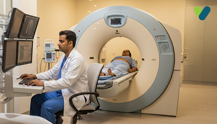
A heart CT scan is used to capture an internal image of the heart and its blood vessels. It uses a strong X-ray machine to obtain images of the heart to find problems like blocked arteries. The test doesn't need any cuts or needles, so it's not painful. The test has no physical implications on your body, so you can return to regular activities soon after the test.
This test can have different names depending on the doctor's preference. If they want to check if there is too much calcium in the tubes, which provide the blood to your heart, it's called a coronary calcium scan. If the test is done to diagnose narrow or blocked arteries, it's called a CT angiography. In this test, the doctor also checks other vessels in your body in combination, like the pulmonary arteries or aorta, to look for any problems with these structures.
Heart CT Scan: Purpose
This test is performed to look for certain heart conditions, including congenital heart disease, primary valve injury, tumour, blood clots or hard substance build-up. Your doctor may suggest a cardiac CT scan and determine what is wrong. A heart CT scan is a common test that can help your doctor see the structure of your heart and its blood vessels without any cuts or needles.Possible Complications of a Heart CT Scan
This procedure is usually safe and doesn't have many risks. However, there can also be some risks, like those with any regular scan. This may include radiation exposure, allergies to the contrast dye used in some CT scans, and kidney problems, especially if you have an existing condition.Moreover, people having severe asthma should take the test under the supervision of a physician. This is because the dye can make breathing difficult for people with severe asthma or cause their airways to narrow.
Even though the amount of radiation received from a heart scan is low, you should tell your doctor if you are pregnant or make sure they know about your allergies or kidney problems.
How to prepare for the test?
Before a heart CT scan, it is important to follow the instructions that your healthcare provider or the imaging centre staff demonstrates. However, there are some common guidelines that everyone should know about before taking this test:- Inform your healthcare expert about your medical conditions, such as kidney problems or allergies, if any. Furthermore, if you are pregnant or think you might be, you should convey it to them.
- If you are given specific instructions about food and drink restrictions, follow them strictly. Your instructor may ask you to avoid eating or drinking for a few hours before the scan.
- Always wear comfortable, loose-fitting clothes when taking this test. Ensure that your clothing has no metal fasteners or accessories, as these may interfere with the process.
- If you are receiving a contrast dye injection, you should have a blood test before the scan to check your kidney function, and if you have a history of contrast dye allergies, inform your healthcare provider.
- Arrive at the imaging centre on time. Also, you can ask the staff any concerns or questions regarding the test.
What Can You Expect During the Procedure?
The process is conducted by the radiology department in an outpatient imaging facility or a special clinic. Here is what to expect before, during and after the test.Before the Test
Sometimes, you might need to take beta-blockers, a medicine, before the scan. This medicine helps to take better pictures by slowing down your heartbeat.
The technician will place sticky discs, which are tiny, termed electrodes on your chest for the scanning process. They will also put a needle into the vein to inject the contrast material into the arm. You might have a metallic taste in your mouth or feel flushed or warm for a short time when the dye is injected.
Before the scan starts, you will lie down on a table, and sometimes you must lie in a certain position. The technician might use straps or pillows to help you remain in the right position to acquire a good picture.
During the Test
During a heart CT scan, you will lie down on a table that moves into a big CT scanner machine. The machine takes many X-ray pictures of your heart and blood vessels from different angles. A computer combines these pictures to make a 3D image of your heart
Sometimes, you may receive a special dye through an IV in your arm. This dye helps your blood vessels and heart appear more clearly on the scan.
The scan usually takes 10–15 minutes, and you must stay still to ensure that the pictures are clear. A technician will operate the scanner from another room and talk to you through an intercom.
After the cardiac CT scan, a doctor will look at the pictures to check for any problems. Then, the results will be given to your regular doctor, who will discuss and suggest any needed treatments or follow-up care.
What Happens After the Procedure?
Once your CT coronary angiogram is finished, you can resume your regular daily routine. You can drive yourself back home or to work. Drinking plenty of water helps to remove the dye from your body.Based on the results, your doctor may suggest some changes for you to make your lifestyle better, like eating healthy foods or giving up smoking. It may help to reduce your chances of having heart problems in the future.
It's essential to follow your doctor's advice if they recommend attending the appointments to keep track of your heart healthy.
Results
The images taken during your CT coronary angiogram should be available shortly after your test. The healthcare provider who requested the test will review the results and discuss the findings with you.
If the test shows a positive result for a condition, you can talk to your healthcare expert to improve and reduce the potential risk of heart disease.
To monitor your heart health, visit the doctor regularly so that they can make any changes to your treatment if needed.
Remember, caring for your heart is vital for staying healthy and feeling good. If you make positive lifestyle changes and work closely, as your health expert says, it can help keep your heart healthy and strong.
Ways to Improve Heart Health through Lifestyle Changes
Irrespective of whether any issues are found in the CT coronary angiogram, it's essential to make lifestyle changes to promote a healthy heart. Here are some tips to help you out in improving your lifestyle:- Exercising daily can be beneficial for you to control your weight and manage diabetes, high cholesterol, and high blood pressure. Your healthcare provider can guide you on how much exercise you need.
- Smoking is another reason that can cause heart disease, and stopping smoking is the best way to lower this risk. You can ask your doctor for assistance in quitting.
- Eating nutritious food such as fruits, vegetables, and whole grains and avoiding fatty and unhealthy foods may benefit you to make this condition better.
- In case you are diagnosed with such health conditions, take your prescribed medicine and see your doctor as often as they suggest.
- Stress can increase your risk of a heart attack. Some ways to manage stress include regular exercise, practising mindfulness, and seeking support from close ones.
A heart CT scan is a non-invasive procedure that has minimal risks and requires little preparation. By following the instructions provided by healthcare professionals, patients can ensure accurate results and a comfortable experience during the scan.
After the scan, you can resume your daily activities. If the presence of heart disease or other conditions is diagnosed in the scan result, you can talk to your healthcare adviser about treatments and ways to make you healthier.

