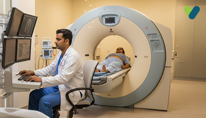
MRI pelvis Also known as a pelvic MRI scan, an MRI pelvis is a pain-free procedure that is a preferred imaging test for pregnant women and young children. The MRI pelvis uses magnetism and radio waves to create a detailed image of the internal organs of the pelvic region (which is the area between the hips), including the bones, soft tissues, and blood vessels. The pelvic region contains the bladder, reproductive organs, large intestine, small intestine, pelvic bones, and prostate, all of which can be seen clearly in an MRI pelvis.
This imaging test is preferred over other tests, such as CT scans or X-rays, as it does not use radiation, which makes it a safe procedure, and it produces images with more details. Using an MRI pelvis test helps the doctor make a quick diagnosis without the need for exploratory surgery or cutting the body to see the insides for any abnormalities. The MRI pelvis test is conducted in a secluded room where only the patient remains inside the machine due to the presence of a strong magnetic field. The MRI machine looks like a large hollow tube with a table in the middle that slides in and out of it.
Why is an MRI pelvis test recommended?
A doctor may recommend a pelvic MRI if the patient has any of the following:- Congenital or birth defects in the pelvic region
- Injury or trauma to the pelvic region
- Pain in the lower abdomen or pelvic region
- Unexplained problems in urination (peeing) or defecation (pooping)
- Cancer or the probability of cancer in the bladder, rectum, urinary tract, or reproductive organs
- Abnormalities in other test results, such as X-rays
- Infertility
- Irregular vaginal bleeding
- Unexplained pain in the pelvic region
- Masses or lumps in the pelvic region, such as uterine fibroids
The doctor usually shares the reason for ordering an MRI of the pelvis during the patient’s visit, but the patients are encouraged to understand the reason and outcomes of the test before going through with it.
Preparing for the exam
The doctor usually asks the patient about any metallic implants or other medical devices in their body, as well as any allergies they may have. However, patients should proactively mention these details and clarify any doubts before the procedure. The MRI machine uses magnetism during the scan, which can be extremely harmful if the patient has a metal object on them or inside them. Metal inside the body may move, causing internal bleeding and/or becoming heated.People who have a fear of confined spaces are recommended to take medication to calm their nerves before the procedure, as the machine may induce claustrophobia. Patients must not eat or drink anything up to four hours before the scan and follow other guidelines provided by the doctor. The healthcare provider will ask the patient to empty their bladder about two hours before the scan and not go to the restroom until the scan is complete, which can take anywhere from 30 to 60 minutes. It is advisable to bring a friend or family member for support.
Pelvis MRI scan procedure
The test may last up to 60 minutes or more if the results are not clear enough on the first attempt. The procedure usually starts with the patient going through a preparatory checklist with a healthcare provider who operates the MRI machine. If necessary, the patient may change into a hospital gown. Then, the patient is asked to lie flat on their back with their feet facing the machine on the examination table.Depending on the type of pelvic MRI, the healthcare provider may place a comfortable coil around the patient's hips and/or administer a contrast dye to enhance image clarity. In some cases, the healthcare provider may also insert a probe into the patient's rectum if the test requires it.
Once everything is in place, the healthcare provider will leave the room after showing the patient how to use the two-way intercom. They operate the machine from a different room, and the machine starts making loud humming, clicking, and thumping sounds. The patient can ask for headphones or earplugs to help remain calm and still during the process.
Associated risks
There are not any major risks associated with a pelvis MRI scan as it does not use radiation. However, people who have certain metal implants (e.g., metallic pins) or medical devices (e.g., a pacemaker) in their bodies should not get an MRI. The patient must inform the doctor beforehand if they have any of the following:- Artificial joints or heart valves
- Metal screws, plates, or pins from previous surgeries
- Pacemaker
- Bullet or its pieces
- First-trimester of the pregnancy

