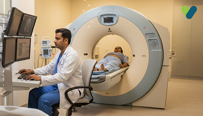
The full form of PSMA PET is prostate-specific membrane antigen positron emission tomography. Prostate cancer detection and treatment are substantially enhanced by the novel prostate-specific membrane antigen (PSMA) PET imaging. The medicine is FDA-approved and is used in the positron emission tomography (PET) imaging of PSMA-positive lesions in patients with prostate cancer. In order to assist in pinpointing prostate cancer cells, the radioactive imaging agent 68Ga-PSMA-11 attaches to prostate cancer cells.
Definition of PSMA PET Scan
Prostate cancer can be found throughout the body by using an imaging test called a prostate-specific membrane antigen positron emission tomography (PSMA PET) scan. It employs a radioactive material that seeks a protein known as PSMA, or prostate-specific membrane antigen, which prostate cancer expresses. It is more reliable for detecting prostate cancer than most other imaging tests.How long does a PSMA scan take?
Although scheduling can differ, the PSMA PET scan typically proceeds for around 2 hours. A nurse or technician will administer a special dye with a radioactive tracer into one of your veins to do a PSMA PET scan. To allow the dye to spread throughout your body, they will ask you to wait for 30 to 60 minutes. Further, you will have to lie down on a cushioned exam table. To create images of your body, they will pass the table through a PSMA PET CT scan or PET-MRI scanner. This scan takes 30 minutes to complete the whole process.You will get the results within a timeline of 1 to 2 weeks. A professional will examine the images after the scan is complete and give their findings to your doctor. Then, the doctor will discuss the findings with you to deduce a treatment procedure.
What Happens During a PSMA PET Scan?
The radioactive agent that targets PSMA is injected by a technician into a vein in the patient's arm. The patient is placed on a table that glides in and out of a doughnut-shaped machine an hour later. The device gathers several images of the body's inside organs. To get more precise images, the scan is performed using either computed tomography (CT scan) or a magnetic resonance imaging (MRI) scan. The photos are all reviewed by a radiologist, who notes any bodily parts with high PSMA levels, such as prostate cancer.Is there any risk factor during PSMA PET Scan?
For most patients, PSMA PET scans are safe. Some people may notice transient symptoms, including weariness, taste changes, or headaches that will go away on their own. There is a small chance of an allergic reaction, especially in patients with a history of allergies to other drugs and foods. The scan also raises a patient's overall radiation exposure, which may slightly raise their risk of developing cancer in later life.Limitations of the PSMA PET Scan
Due to the small percentage of prostate tumours that do not express PSMA and are therefore undetectable by a PSMA PET scan, there is a slight possibility of a missed diagnosis. Sometimes, PSMA is expressed by non-cancerous diseases and by other cancers, leading to inaccurate diagnoses of prostate cancer. Despite being uncommon, a missed or inaccurate diagnosis may prompt doctors to prescribe the wrong course of action.Use of a PSMA PET scan
The following circumstances necessitate the use of a PSMA PET scan:- To eliminate the risk of cancer spreading from the pelvis to other regions of the body in men with prostate cancer.
- To establish a patient's eligibility for the radiopharmaceutical treatment lutetium Lu-177 vipivotide tetraxetan, which is used to treat prostate cancer that expresses PSMA. Patients with prostate cancer who have advanced during chemotherapy and other cutting-edge oral hormone treatments may benefit from this scan.
- Prostate-specific antigen levels may rise in patients who have previously undergone radiation or surgery for prostate cancer. Physicians and patients may choose between systemic therapy and radiotherapy based on the findings of a PSMA PET scan.
Why should you choose a PSMA PET scan for prostate cancer?
It is quite difficult to locate prostate cancer with sufficient accuracy for treatment because in cases where the tumours have migrated outside of the prostate, prostate cancer tumours can be identified throughout the pelvis and the body.Prostate cancer is the second most prevalent disease in males and the second largest cause of cancer-related death in men. Hence, it is crucial to accurately locate and remove these tumours.

