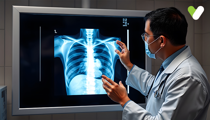
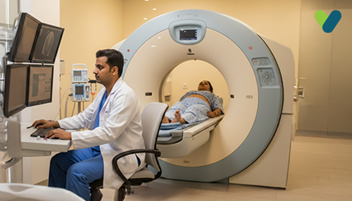
What is a digital X-ray?
A quick digital radiographic picture is obtained using an advanced X-ray imaging method called digital X-ray/digital radiography (DR). With this method, the data can be gathered when an object is being studied with X-ray-sensitive plates, and the data is then uploaded to a computer system without the need for an intervening medium without delay. A detector sensor transforms the incident X-ray beam into a comparable electric charge before turning it into a digital picture.Digital radiography requires the use of an electronic image capture system rather than an X-ray film. The benefits of digital X-rays include quick image preview and accessibility, the removal of expensive film processing procedures, a dynamic window, and access to specialised methods for image processing that improve the overall visual appeal of the image.
Uses of digital X-ray
Digital X-rays help the physician inspect a patient's body. This could help determine the degree of damage caused by an injury, such as the dislocations and fractures of bones. They can also spot soft tissue lumps that might indicate the presence of cancer or other diseases.Dental professionals are particularly interested in digital X-ray because it provides rapid access to the results, which allows them to improve the images by adjusting their exposure in real-time and, as a result, receive detailed and clear reports that can be instantly shared with their patients. Digital X-rays are better than conventional X-rays in terms of detecting minute fractures and deformities in the teeth because of their level of detail.
Advantages of digital X-ray
Real-time enhancement: The potential to produce the images instantly allows the doctor to change the brightness in real-time, obtaining darker or lighter images as necessary to enhance the visibility of specific scan components.Reduced radiation exposure: Digital X-rays create high-quality pictures immediately with less radiation exposure than traditional X-rays, which proves beneficial for the patient. Clarity: A physician or dentist can see the scanned region of the human body in exceptionally fine detail due to the high-quality pictures that digital X-rays can produce. Speed: It is possible to review your results instantaneously with the help of a digital X-ray test.
Reduced chemical use: In a digital X-ray test, the use of chemicals to produce high-quality images is avoided. Efficiency: In addition to using less radiation, digital X-rays save a plethora of effort and time compared with conventional X-rays because the results are instantly and readily accessible with the click of a button.
Limitation of digital X-ray
- Equipment for digital radiography is very costly because tools such as computers, servers, and recording devices are needed.
- Depending on the size of the images, the images need considerable storage space.
- Digital pictures can be intentionally altered for any purpose.
- In comparison to a radiographic film, most storage phosphor technologies offer a poor optimal resolution.


