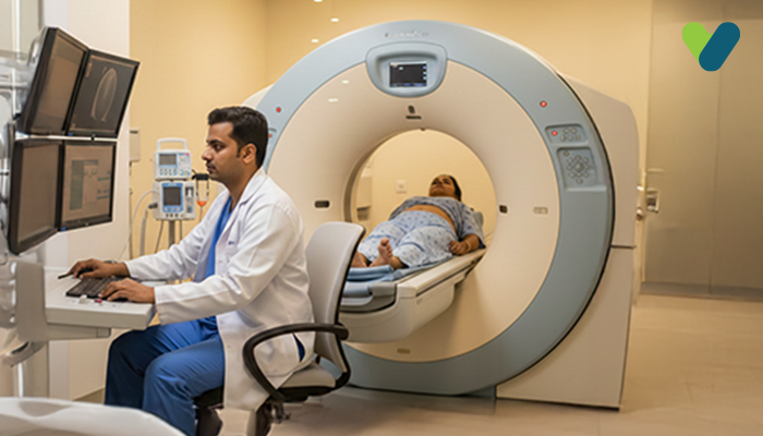
Computed tomography, also known as a CT scan or CAT scan, is a diagnostic procedure that generates the pictures of the interior of a body with the help of computer technology and X-rays. It displays detailed photos of various parts of the human body, such as bones, muscles, organs, and blood vessels.
CT scans provide more information than standard X-rays. The X-ray beam in a CT scan moves in a circular pattern around the body. This allows for multiple views of a particular structure or organ and provides considerably more details. The X-ray information goes to a computer, which interprets it and displays it on a monitor in a two-dimensional form. Nowadays, with advanced technology, three-dimensional imaging is also possible.
How are CT scans performed?
A CT scan can be performed at a radiology clinic or hospital. Your doctor may instruct you to fast for a couple of hours prior to the procedure. Wearing a hospital robe and removing any metallic objects, including jewellery, belts, dentures, or eyeglasses, may also be required.A radiology technician will perform the CT scan. While performing the test, you will have to lie down on a table in a big, ‘doughnut-shaped’ machine. Just as the table begins to move slowly through the CT scanner, the X-rays go around your body. Hearing a buzzing or whirring noise during the procedure is absolutely normal. Since any movement during the scanning may result in blurring of the image, you will be instructed to remain perfectly still. You may also need to hold the breath at times.
The duration of the CT scan is determined by the body part being scanned. It may only require somewhere between 10 and 30 minutes, or even less, to complete the scanning. In almost all cases, you will be able to go back home the very same day.
Why are CT scans performed?
A CT scan can be used for various reasons, which include:- To help detect joint and bone issues such as tumours and complex fractures.
- To identify or help doctors see any changes if you are suffering from disorders such as cardiovascular disease, cancer, liver masses, or emphysema.
- Doctors can make comparisons of CT scans to determine whether a specific treatment is effective. For instance, the CT scans of a tumour over time can tell whether it is responding to radiation therapy or chemotherapy.
- They help doctors guide treatment plans and procedures, including biopsies, radiation therapy, and surgeries.
- CT scans can show internal bleeding and injuries, for example, those caused in a car accident.
- They can aid in the detection of excess fluid, a tumour, a blood clot, or an infection.
What is a CT scan with contrast?
A CT scan can clearly show dense substances such as bones. On the contrary, soft tissues are hard to see and may appear blurred in the image. To make them stand out, a special material or dye referred to as contrast media might be required. These dyes absorb X-rays and show up in white colour on the scans, thus highlighting organs, blood vessels, as well as other structures.Barium sulphate and iodine are the two common contrast materials used. You could get these drugs in any of the following three ways:
- Oral: Consuming a liquid containing a contrast medium can improve the scans of the digestive tract, which serves as the pathway for food through the body.
- Injection: The drugs are administered intravenously. This is done in order to improve the visibility of your liver, urinary tract, gallbladder, or blood vessels in the image.
- Enema: The contrast material can be inserted into your rectum if the intestine is being scanned.
You will need to consume a lot of fluids after the CT scan to help the kidneys eliminate the contrast dye from the body’s system.
Are there any risks associated with a CT scan?
CT scans have been using X-rays, which produce ionising radiation. As per the research, this type of radiation can damage your DNA and even cause cancer; however, the risk remains insignificant.Even so, the effects of radiation continue to accumulate with each scan. Therefore, your risk goes up with each CT scan. You should ask your doctor about the procedure's potential risks and benefits and inquire about the reason why a CT scan is required.
In children, radiation exposure may be more dangerous than that in adults because they are still growing. Prior to the procedure, you should check with the physician or technician to see if the CT scan machine's settings have been adjusted for a child.
In some cases, the doctor may advise you using a special dye known as contrast material. Although it is uncommon, the contrast material or dye can cause medical issues or allergic reactions. Most of the reactions are mild, resulting in itchiness or a rash. An allergic reaction could be serious, even fatal, in rare cases. Inform your doctor if you have ever had an adverse reaction to contrast media in the past.
Let your physician know just in case you are pregnant. Despite the fact that the radiation from the CT scan is unlikely to harm your unborn baby, the doctor may recommend you to undergo some other type of test, for example, an MRI or ultrasound, to prevent exposure of your baby to radiation.
Takeaway
CT scans are a great diagnostic tool for soft tissue, blood vessels, and other body part problems that cannot be seen with ultrasound or X-ray imaging.These painless scans require little preparation and may be performed quickly in an emergency. A CT scan requires just under an hour to complete, but results may not be available immediately, based on the individual interpreting the results.
After evaluating the images, your doctor will determine whether a contrast substance is necessary for performing the scan as well as the action you should take.

