Livlong 365
HomeLab TestLiver Ultrasound: Overview, Uses & Preparation

Liver Ultrasound: Overview, Uses & Preparation

Overview of Liver Ultrasound

An ultrasonic scan uses sound waves with high frequencies to take real-time pictures of your organs. The ultrasonic scan is a less invasive option than some other procedures. A doctor uses liver ultrasound, also known as liver sonography, to diagnose the liver by sending sound waves with high frequencies into your liver. The waves often rebound from the inside of your liver and produce an image or video that may be seen on a monitor.
Examining the liver’s normal structure and abnormalities becomes very easy using liver ultrasound. The structure, masses, and dark or light-coloured lesions observed during liver ultrasound give a doctor information regarding several liver illnesses, including fatty liver disease, cysts, cirrhosis, and hepatitis.

Liver ultrasound is a vital tool that gives medical professionals a real-time view of the liver and its arteries. It is a specific kind of abdominal ultrasound imaging technique. If your doctor has requested liver ultrasound exam for your, they may be looking for verifying or determining a liver problem.

Uses of liver ultrasound

Liver ultrasound can provide essential data regarding any liver abnormalities. To find hepatitis, cysts, cirrhosis, fatty liver, and other conditions, doctors look at the density and brightness of the liver sonography. Cysts and solidified masses can be easily distinguished through liver ultrasound scan. Simple cysts are made up of fluids and a thin outer layer, and they appear as dark masses on liver ultrasound scans. Nodules, lumps, calcifications, or many tissue layers are frequently found in complex cysts. Doctors may be able to tell if you had complex cysts from the solid portions (nodules and lumps). Distinct liver disorders, such as cirrhosis, hepatitis, and fatty liver, can also be detected by liver ultrasound. The scan typically shows a fatty liver to be brighter than a healthy liver. The liver scan appears less vibrant when hepatitis is the underlying issue.

Many medical professionals advise the use of liver ultrasound and elastography to evaluate the liver’s flexibility. It may additionally be utilised to determine the probability of fibrosis. Liver ultrasound can also reveal enlarged bile canals and fluid in the nearby areas of your liver. Moreover, liver ultrasound frequently reveals the details of a few organs, including the gallbladder, right kidney, and a small section of the pancreas. Because of the differences in appearance between malignant and non-malignant liver tumours, liver ultrasound can help detect liver cancer.

For several reasons, doctors typically advise liver ultrasound to be performed:

  • If you experience discomfort in the upper portion of your abdomen where your liver is located or show some other signs of liver illness like jaundice
  • If your liver enzyme levels are elevated
  • For the preparation and monitoring of liver transplants
  • For monitoring the results of liver cancer therapy
  • For monitoring the results of TIPS (transjugular intrahepatic portosystemic shunt) a surgery used in individuals with liver cirrhosis
  • For identifying obstructions (clots) that lead to decreased blood circulation in the veins and arteries within and surrounding the spleen and liver

Additional uses of liver ultrasound:

To obtain a more precise image and identify potential problems, medical professionals can pair liver ultrasound with other methods. The following are some typical liver ultrasound exams:

Elastography: This method is used to examine the stiffness of the liver tissue, which may indicate cirrhosis or another disease. To view the liver’s tissue, a sequence of impulses needs to be sent to the liver.

Contrast imaging: To provide clear visuals of the liver and its blood vessels, this procedure includes inserting dye inside the blood vessels. It can be particularly useful for identifying liver cancer.

Combined techniques: To obtain highly precise images of the interior of the liver, a physician could combine different techniques, such as magnetic resonance imaging (MRI) and liver ultrasound.

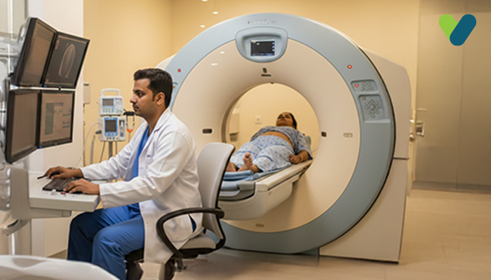
What to expect from liver ultrasound?

Your doctor will advise that you avoid fatty foods for several hours before liver ultrasound since the liver has a significant function in the digestion of fats. Because liver ultrasound is not painful and is a safe exam, it can be done as an outpatient procedure. If you have any queries or worries, your technician can guide you through the process beforehand. You may feel a tiny chill as a gel comprising water is gently applied to your abdomen. The gel guarantees that there is a firm connection between the layer of skin and the probe (instrument), ensuring that the sound waves travel straight to the tissue. To get the most accurate reading, the technician may frequently ask you to change positions. It normally takes thirty minutes to finish the entire liver ultrasound; however, this time may increase based on the size of the organ. No recovery time is needed after liver ultrasound. Following the scan, an individual can consume food, beverages, drive, and go back to their job or school.

Preparations for liver ultrasound

Preparation is not always necessary for liver ultrasounds. Anaesthesia and painkillers are not required for liver ultrasound since this procedure is safe and non-invasive. For liver ultrasound, a doctor might recommend the patient to fast before the scan. This is because the gallbladder may expand during fasting, making it easier to view.

Risks of liver ultrasound

Liver ultrasound rarely involves any genuine risks. Since the examination is safe and has no clear absolute limitations, almost anyone is eligible for it. However, if a woman is pregnant, doctors typically advise against using pulsed Doppler methods directly over the foetus. When liver ultrasound is used with other examinations, adverse effects are possible. For instance, injecting contrast dye could harm, and in exceptionally rare circumstances, someone could experience an allergic response to the dye.

Dr.William Lewis Aliquam sit amet dignissim ligula, eget sodales orci. Etiam vehicula est ligula, laoreet porttitor diam congue eget. Cras vestibulum id nisl eu luctus. In malesuada tortor magna, vel tincidunt augue fringilla eget. Fusce ac lectus nec tellus malesuada pretium.

MBBS (Bachelor of Medicine & Bachelor of Surgery) Gold Medalist (2009-2015) M.D In General Medicine (2016-2019), CCID (Infectious Diseases)

PG Diploma In Clinical Endocrinology v& Diabetes, Clinical Associate in Non-Invasive Cardiology

Relevant Articles

Blog

CECT Abdomen Scan: Purpose,...

What is a CECT abdominal scan? Contrast-enhanced (CE) computed tomography (CT),...

PSP Test: Overview, Procedure...

The phenolsulfonphthalein (PSP) test was introduced in 1911 as a...

Subscribe Today

Receive Meaningful & Informative Blogs directly in your inbox

How to Gain Weight : Expert Weight Gain Diet Plan & Tips

0
Lalita, 23, has always been a skinny girl. Ever since her childhood, Lalita has been at the receiving end of constant ridicule because of...

Spondylosis : Exploring the Causes, Symptoms, Treatment, Types

0
Spondylosis is a general medical term for the wear and tear of the spinal disc and joints. This is a common cause that affect...

Tinnitus: Ringing in the ears and what to do about it

0
Imagine sitting in a quiet room, but there is this ringing or buzzing that won't let go of your ears. Such was the case...

Frozen Shoulder: Thawing the Painful Condition of Adhesive Capsulitis

0
Mark, a 45-year-old IT professional could not fathom why his left shoulder had stiffened and caused pain for a few months. Even reaching into...