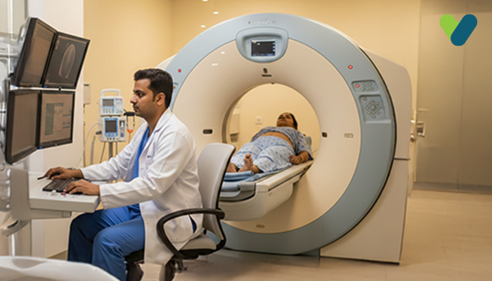
Breast cancer is one of the most prevalent types of cancer, yet it is highly curable when detected in its early stages. A mammogram is a screening as well as a diagnostic tool used for detecting any changes related to cancer that occur in the breast tissue even before symptoms appear.
Here, we will discuss what a mammogram is, how it works, how to prepare for one, and what to expect during mammography.
What is a mammography test?
Mammography is a type of medical imaging that employs a low-dose X-ray system for examining the inside of one's breasts. A mammography exam, also referred to as a mammogram, helps identify and diagnose breast disorders early on.It can be used as either a screening tool for breast cancer or as a diagnostic tool; diagnosis might include investigating unusual findings on the other imaging test or any concerning symptoms.
During mammography, the breasts are pressed between two hard surfaces in order to spread out the breast tissue. Then, an X-ray generates black-and-white pictures that are presented on a monitor and assessed for the signs of malignancies.
Traditional mammography produces two-dimensional pictures of the breast. New developments in this technique include breast tomosynthesis, which produces three-dimensional images of the breast.
How do mammograms work?
X-rays are a type of radiation that can pass through most objects, including the human body, similar to light or radio waves. An X-ray technologist aims the X-ray beam at the specific area of interest. A small burst of radiation is produced by the machine, which then passes through the body and creates an image on a specialized detector or photographic film.X-rays are absorbed by different areas of the body at varying rates. Soft tissues like fats, muscles, and organs let more X-rays pass through, while dense bones absorb a significant portion of the radiation. Consequently, the soft tissue appears in shades of grey, while bones are visible in white color on an X-ray image. Air, on the other hand, appears black on an X-ray.
The majority of X-ray pictures are saved electronically. Your doctor will have easy access to these saved images to determine and manage the condition. In conventional as well as digital mammography, a stationary X-ray tube provides an image from above the compressed breast and a picture from the side. The X-ray tube travels in an arc across the breast during breast tomosynthesis, taking several photos from various angles.
Why is a breast mammogram performed?
Mammograms are X-ray pictures of your breasts that are used to identify cancer and other abnormalities in the breast tissue. Mammography can be used for either screening or diagnosis.- A screening mammogram is a type of breast cancer screening tool used to identify potential cancerous changes in the breasts of people who do not have any symptoms. The purpose of this type of screening is to detect cancer in its early stages when it is small and easy to treat.
- Diagnostic mammography is performed for the investigation of any concerning breast changes, such as breast soreness, a new breast lump, abnormal skin appearance, nipple discharge, or nipple thickening. It is also used to assess abnormal findings seen in screening mammography. During a diagnostic mammogram, additional mammogram images are taken to investigate the abnormalities further.
When should you get a mammogram?
According to 'The American College of Physicians' guidelines, the following screening plan is recommended for women with an average risk of developing breast cancer:- For individuals between the ages of 40 and 49 years, it is recommended that they seek guidance from a doctor regarding mammography screening.
- For those aged 50–75 years, it is generally advised to undergo mammography screening every two years.
- Once an individual reaches 75 years of age, it is usually recommended to discontinue mammography screening. People who have any or all of the following conditions might need additional screening:
- Genetic factors, including BRCA1 or BRCA2 gene mutations
- High-risk lesions in the breast or personal history of breast tumor
- A history of childhood chest radiation exposure
How to prepare for mammography?
When planning for your mammography, keep the following points in mind:- Inform your healthcare practitioner if you are pregnant, breastfeeding, or suspect you might be pregnant. In such a scenario, they might advise a breast ultrasound instead of a breast mammography.
- Avoid scheduling your mammography a week before or during your menstrual cycle. This is because your breasts may be sore during this time, making the procedure more unpleasant.
- Please let the examiner know if you have implants in your breasts or if you have recently received a vaccination. On the day of mammography:
- Continue following your usual schedule, including eating, drinking, and taking medications.
- Carry all your previous mammograms with you. This will help the radiologist compare the previous results with the new ones.
- Wearing perfume, deodorant, body powder, or lotion can interfere with the accuracy of the X-ray pictures; therefore, these should all be avoided.
- Some women prefer to put on a top and bottoms rather than a dress. For the imaging procedure, you will need to take off your clothes from the waist up. You will be required to wear a medical drape or gown.
What to expect during a mammography procedure?
The following steps are involved in mammography:- Any clothing and jewelry above your waist must be removed. You will be given a front-open hospital robe or gown to put on.
- For the mammography procedure, a qualified radiologic technologist will position you in front of a mammography machine. A technician then positions one of the breasts on the specialized platform and adjusts its height to match yours. Your head, torso, and arms are all placed to provide a clear view of the breast.
- Then, the technician presses down your breast against the platform with the help of a transparent plastic paddle. Gradual pressure will be applied for a couple of seconds to spread out the breast tissue. During this time of compression, you might experience some pressure or discomfort. If you are unable to endure the pressure, tell the technician, and they will adjust the instrument accordingly.
-
- To even out the thickness of the breast to ensure that the entire breast tissue could be seen
- To spread the breast tissue out so that the small abnormalities are unobstructed by surrounding tissue
- To allow for the use of a lesser X-ray dosage since the breast tissue being scanned is thinner
- To hold the breast unmoved to minimise image blurring due to movements To minimise X-ray scattering to improve image clarity.
- You will be instructed to switch between photos. A top-to-bottom view plus an angled side view are the standard viewpoints. The procedure will be repeated on the other breast.
- The procedure will be completed after the technician has finished taking X-rays.
- You can put your clothes back on and resume your normal routine. From start to finish, a mammogram takes roughly 20 minutes.
Does mammography hurt?
Given that compression is applied to your breast tissue while performing mammography, some people feel uncomfortable. The bright side is that mammography is quick, and the discomfort does not last long. Inform the technician right away if you are experiencing severe discomfort.The intensity of pain you may experience is determined by a number of factors, including:
- Your breasts' density as well as size
- The amount of compression your breasts require
- If you are about to start or are currently in your menstrual cycle
- The radiology technologist's skills
- Your ability to keep calm
Is the mammography procedure safe?
Mammography subjects your breasts to low levels of radiation; however, the advantages outweigh any potential risk from the resulting radiation exposure. Inform your healthcare practitioner and technologist if you suspect you are pregnant. Although mammography is typically safe to perform during pregnancy, healthcare experts normally advise deferring mammograms for screening purposes if you do not have a high risk of getting breast cancer.Does mammography interfere with breast implants?
Having saline or silicone breast implants, as well as the formed scar tissue, makes it harder for radiologists to view all of your breast tissue and any potential abnormalities on routine mammograms.People with implants frequently have two extra images taken of each of their breasts in addition to the four regular images obtained during screening mammography in order to help the radiologist view as much of the breast tissue as possible. These additional images are known as implant displacement views.
For the implant displacement view, the breast implant is pushed towards the chest wall by the technologist, who then pulls the breast forward over it. This step is followed by compressing the breast to enhance the imaging of the front part of each breast.
When scheduling your mammogram, it is important to notify the mammography facility that you have breast implants and inform the technician on the day of your mammogram.
Who interprets the findings of mammography and how to get them?
The pictures will be analysed by a radiologist, a doctor who is qualified to supervise and interpret radiology examinations. The radiologist will provide you with a signed report, which you will review with your general physician or referring physician.The facility in which you undergo mammography will also notify you of the results. Sometimes, a follow-up exam may be required. If this is the case, your healthcare provider will explain the reason. At times, a follow-up examination may be necessary to further analyse a suspected problem with additional viewpoints or specific imaging technology. It may also be done to check whether a problem has changed over time. Follow-up assessments tend to be the best approach to determining if therapy is effective or whether a condition requires addressing.

