An MRI (magnetic resonance imaging) of the lumbar spine is a painless diagnostic procedure that enables a doctor to examine the insides of the lower section of the spine—the lumbosacral spine. Using magnetism and radio waves, an MRI of the lumbar spine creates the detailed image of the lower back.
The MRI lumbar spine scan examines the five lumbar vertebrae (L1 to L5), the sacrum (the shield at the bottom of the spine), and the coccyx or tailbone. The doctor can see parts, such as the bones, disks, spinal cord, nerves, tendons, blood vessels, ligaments, and cartilage, with the help of a lumbar MRI scan. This type of MRI is helpful in diagnosing different health conditions affecting (or originating from) the lower back region where the lumbar spine is located.
Since this test does not use radiation, it is a safe option for people who can’t have X-rays or a CT scan, such as young children and pregnant women. Please note that women in their first trimester of pregnancy are usually not recommended an MRI unless it’s absolutely necessary. Depending on the patient and their condition, the doctor may suggest a contrast lumbar MRI scan. This contrast MRI uses a safe and special dye, which is administered to the patient intravenously, and some people may develop an allergic reaction to it.
Why is an MRI lumbar spine recommended?
Doctors use this test to diagnose and treat a patient who has problems with their spine, especially the lower back region. The problems can originate from pain related to a past or recent injury, disease/infection, or other factors that can be diagnosed with the help of an MRI lumbar spine scan. Patients who have any combination of the following symptoms are generally recommended an MRI lumbar spine:- Fever accompanying back pain
- Congenital or birth defects impacting the spine
- Trauma- or sports-related injury to the lumbar spine
- Chronic or severe lower back pain
- Trouble in thinking clearly, learning, and planning
- Stiffness, weakness, and muscle spasms
- Numbness and tingling in the lower spine
- Leg pain
- Problems with bowel and bladder control
Preparation for an MRI lumbar spine
The patient should avoid wearing anything made of or that consists of metal, including jewellery, eyeglasses, and/or jeans with zippers. Metal objects can disrupt the test results and can be harmful to the patient’s safety and health. The MRI room has a strong magnetic field when the machine is running. The MRI machine appears like a large hollow tube with a sliding table in the middle.This may cause anxiety and discomfort to people who are claustrophobic. Thus, patients are recommended to inform their healthcare provider about any fear of tight spaces. During the procedure, the MRI machine makes loud thumping, clicking, and humming sounds that may be uncomfortable for the patient. They can request the healthcare providers for earplugs or headphones.
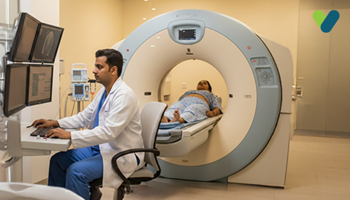
MRI lumbar spine scan procedure
A healthcare provider prepares the patient by helping them change into a hospital gown (if necessary) and positioning them head-first on the table. They will then explain basic guidelines to the patient and place a special coil over the lower body to help get clear imaging. Depending on the type of MRI, the healthcare provider may also administer the contrast dye to the patient intravenously.The table is then pushed inside the hollow tube, and the healthcare provider leaves the room. They operate the machine from an adjacent room with clear windows observing the patient throughout the procedure, which can last anywhere from 30 to 60 minutes.
Once they get satisfactory images, they will conclude the process and help the patient out of the MRI machine. A radiologist usually analyses the MRI results and attaches a report based on their analysis before sending it to the concerned doctor or handing it over to the patient.