HomeLab TestScrotal Ultrasound: Overview, Purpose & Preparation

Scrotal Ultrasound: Overview, Purpose & Preparation

The scrotum is the skin pouch situated at the base of the penis and has the testicles inside it. The organs of the reproductive system of males that generate sperm as well as the hormone testosterone are referred to as the testicles. They are found in the scrotum, in addition to other small organs, the vas deferens, a small tube, and blood vessels.
This page will discuss the scrotal ultrasound, why it is done, how to prepare for the ultrasound, and what happens during the ultrasound.

What is a scrotal ultrasound?

A scrotal ultrasound scan, also known as a testicular ultrasound scan, uses sound waves to create the pictures of the scrotum, especially the testicles. Sound waves are sent into the scrotum by an ultrasound machine, and the pictures are visualised on a computer.

Why are scrotal scans performed?

When doctors are concerned about symptoms like scrotal swelling or pain, they will order a scrotal ultrasound.

A scrotal ultrasound scan can reveal:

  • Signs of testicular injury
  • Testicles size
  • Scrotal veins that are unusually swollen (varicocele)
  • Inflammation or infection of the testicle (orchitis) or epididymis (epididymitis)
  • Fluid accumulation around the testicle (hydrocele)
  • A scrotal cyst or tumour
  • An undescended or absent testicle
  • Testicular twisting (testicular torsion)
  • Infertility

How to prepare for a testicular/scrotal ultrasound

No special preparations are needed for the scan. It is not necessary to abstain from eating or drinking anything prior to the appointment.
Before the exam, a patient needs to remove all garments below the waist. The patient may prefer wearing something easy to remove.

Be prepared to remain as still as doable during the ultrasound procedure in order for the ultrasound equipment to produce clear pictures of the testicles.
Medication can rarely be interrupted or stopped prior to a testicular ultrasound. You should, however, consult your doctor for advice on any over-the-counter or prescription medications you use.

What to expect during the scrotal ultrasound?

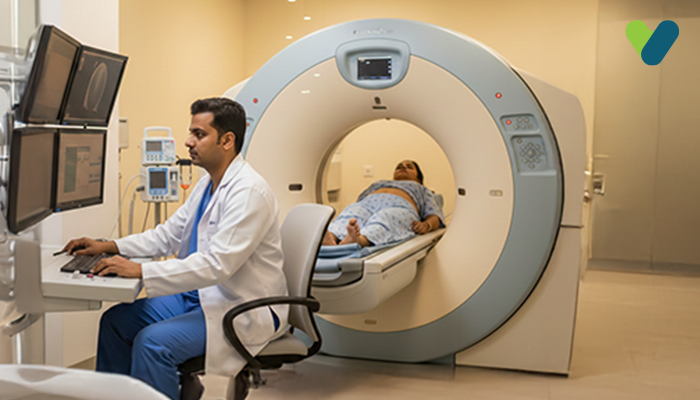
The procedure will take place in your doctor’s office, clinic, or radiology department. It is pain-free and takes about 30 minutes.

You will need to lie flat on the back on the examination table. A water-based gel will be applied to your scrotum by an ultrasound technician. They will move a small tool across your skin (called a transducer) against your scrotum. It does not hurt, but you might feel some pressure.

Sound waves travel from your skin to the transducer. The photos of the testicles will be displayed on the computer monitor. After the ultrasound, the gel will be wiped off, and the photos will be reviewed by a radiologist.

Are there any risks to having a scrotal ultrasound?

A scrotal ultrasound will not put your health at risk. During the procedure, no radiation is used.
Nevertheless, if you are suffering from specific testicular problems, such as an infection or testicular torsion, you may experience increased discomfort or pain during the procedure.

Takeaway

An ultrasound exam is a reliable and efficient method to get a snapshot of what’s going on inside the body.
A scrotal ultrasound should give you a good idea of what is wrong with your testicles. It will assist a physician in recommending additional treatment if necessary.

Dr.William Lewis Aliquam sit amet dignissim ligula, eget sodales orci. Etiam vehicula est ligula, laoreet porttitor diam congue eget. Cras vestibulum id nisl eu luctus. In malesuada tortor magna, vel tincidunt augue fringilla eget. Fusce ac lectus nec tellus malesuada pretium.

MBBS (Bachelor of Medicine & Bachelor of Surgery) Gold Medalist (2009-2015) M.D In General Medicine (2016-2019), CCID (Infectious Diseases)

PG Diploma In Clinical Endocrinology v& Diabetes, Clinical Associate in Non-Invasive Cardiology

Relevant Articles

Blog

CECT Abdomen Scan: Purpose,...

What is a CECT abdominal scan? Contrast-enhanced (CE) computed tomography (CT),...

Five Exercises to Increase...

Many men are often troubled by the length and thickness...

Subscribe Today

Receive Meaningful & Informative Blogs directly in your inbox

Irregular Periods: Causes and How to Manage

0
Does your period sometimes come like clockwork, and other times is it completely unpredictable? Irregular periods can be frustrating and cause confusion or even...

Head-to-Head Differences Between Hallucination and Delusion

0
Hallucinations and delusions both take you to the false reality of life, keeping you struggling with the things that are not real. Both are...

Control Your Blood Pressure: The Complete Guide to Easy Lifestyle

0
Managing blood pressure has been a challenge for most people. Low blood pressure (hypotension) symptoms can include dizziness, lightheadedness, fainting, fatigue, cold clammy skin,...

Acute Renal Failure and Its Impact on Kidney Function

0
Acute renal failure (ARF), also known as acute kidney injury (AKI), is a severe and unexpected disorder in which the kidneys lose their filtering...