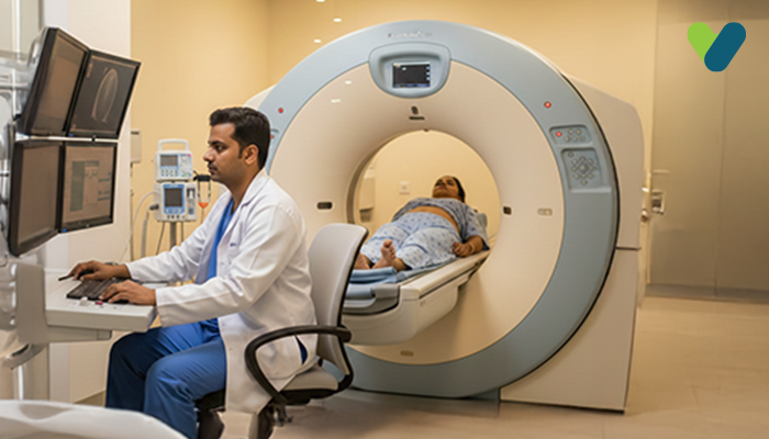
What exactly is a transvaginal (TVS) ultrasound?
An ultrasound test creates the pictures of your internal organs by using high-frequency sound waves. These tests can detect abnormalities and assist doctors in diagnosing various conditions.A transvaginal ultrasound, also known as TVS sonography, or endovaginal ultrasound, is a kind of pelvic ultrasound that doctors use to examine and assess the female reproductive organs, including the uterus, ovaries, cervix, fallopian tubes, and vagina.
The term ‘transvaginal’ refers to ‘through the vagina’. It is an internal investigation. Unlike a standard pelvic or abdominal ultrasound, in which the ultrasound transducer is placed outside your pelvis, this process requires your technician or doctor to insert an ultrasound probe, which is about 2 to 3 inches long, into your vaginal canal.
When should a transvaginal ultrasound be done?
A transvaginal ultrasound may be required for a variety of reasons, including tests for:- Abnormal abdominal or pelvic examination
- Pelvic pain
- Infertility
- Confirming whether an IUD is properly placed
- Vaginal bleeding due to unexplained reasons
- Uterine fibroids or cysts
- Ectopic pregnancy (when the foetus implants outside of the uterus, mostly in the fallopian tube)
- Monitor the foetus's heartbeat closely
- Detect the possibility of a miscarriage
- To confirm an early pregnancy
- Check for changes in the cervix, which might result in complications, including premature delivery or miscarriage
- Check the placenta for any abnormalities
- Determine the root cause of any abnormal bleeding
How to prepare for a transvaginal ultrasound scan?
In almost all instances, a transvaginal ultrasound scan requires little or no preparation on your part. When you arrive at your doctor's office or hospital and enter the exam room, you must remove your clothing from the waist below and put on a gown. Depending on your doctor's instructions and the purpose of the ultrasound scan, your bladder might be required to be partially filled, full, or empty. A full bladder assists in lifting the intestines and provides a clearer view of the pelvic organs.If you are on your period or spotting, you must remove any tampons you are wearing before the ultrasound.
What to expect during a transvaginal ultrasound?
Transvaginal ultrasound is an outpatient procedure and can be done in a hospital, clinic, or radiology department. The whole procedure may take up to 30 minutes. When it is time to start the procedure, you will have to lie on your back with bended knees on the exam table.Your doctor or technician inserts the ultrasound wand (probe) into your vagina after covering it with a condom and lubricant gel. Ensure that your doctor is informed of any allergies to latex you may have in order that a latex-free probe cover can be used if necessary.
You may feel a sensation of pressure when your doctor introduces the transducer. Once the probe is inside your body, waves of sound bounce off your inner organs and transmit images of the interior of your pelvis to a computer monitor. While the transducer is still inside your body, the ultrasound technician or doctor slowly turns it. This gives a complete image of your organs.
Sometimes, a saline infusion sonography may be ordered by your doctor. This is a type of transvaginal ultrasound in which sterile salt water is inserted into the uterus prior to the ultrasound scan to help detect any potential abnormalities within the uterus.
The saline solution slightly stretches the uterus, giving a more comprehensive picture of what's happening inside the uterus instead of a conventional TVS ultrasound.
A transvaginal ultrasound can often be performed on a woman with an infection or a pregnant woman, but saline infusion sonography cannot.
Is it painful to have a TVS ultrasound?
No. The transducer is created to conform to the shape of your vagina in order to make the procedure as pain-free as possible. Furthermore, the lubricating gel on the transducer enables gentle insertion. When the transducer is inserted into your vagina, you might feel some pressure or discomfort.What are the risks of TVS ultrasound procedure?
Transvaginal ultrasound is not associated with any known risks. Transvaginal ultrasounds performed on pregnant women are also safe for both the mother and the foetus as no radiation is used in this technique.You will feel pressure and, in some cases, discomfort while the transducer is being inserted into your vagina. The pain should be minor and subside once the procedure is over. If you experience any discomfort during the exam, please notify the doctor or technician.

