Sonography or ultrasound is an efficient imaging test that can help with the diagnosis of a patient’s illness. Healthcare providers may use an ultrasound to guide a biopsy or surgical procedure. Usually, ultrasounds are considered for pregnancy, but this harmless procedure is used to examine other internal parts of the body as well. Keep reading to learn more.
What is an ultrasound?
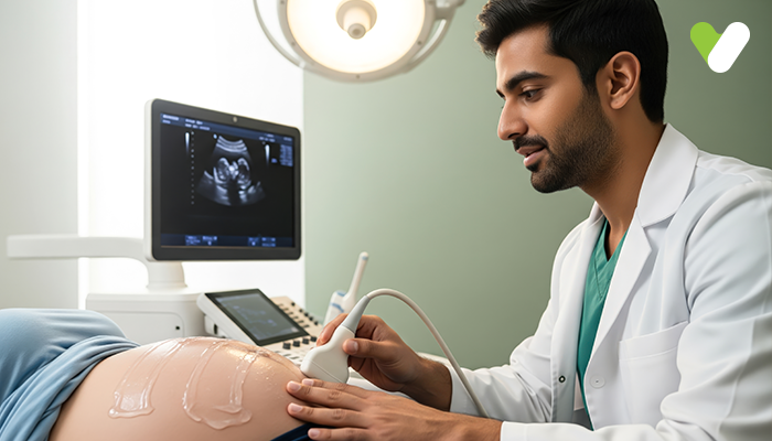
Also referred to as sonography or ultrasonography, ultrasound is an imaging test that helps with diagnoses and treatment. This non-invasive procedure produces an ultrasound picture, which is called a sonogram, using high-frequency sound waves to create real-time visuals (pictures orvideos) of the internal organs or other soft tissues.A healthcare provider uses a transducer or a probe over a part of the body after applying a thin layer of gel to the skin. This transducer converts electrical current into high-frequency sound waves that are inaudible and sends them into your body; the gel helps the waves pass through the skin. These sound waves bounce off the structures inside your body and are caught by the transducer, which then converts them back to electrical signals. Using a computer, the pattern of these electoral signals is converted into real-time images or videos and displayed on the monitor.
This imaging technique enables healthcare providers to see the details of the soft tissues inside the body without making any cuts. This technique is harmless and painless; it does not use radiation like X-rays or CT scans. People associate ultrasounds with pregnancy or checking the health of the foetus, but ultrasound is used for many different situations and procedures in healthcare. Doctors can look at several different parts of the inside of your body using sonography.
Types of ultrasounds
Depending on their purpose, there are three major categories of ultrasounds: Pregnancy/prenatal/obstetric ultrasound Healthcare providers use ultrasound to monitor the foetus during pregnancy. This ultrasound can confirm the pregnancy, check for the number of babies, estimate the gestational age of the foetus or how long the woman has been pregnant, and see the foetus’s position and movement. Additionally, an ultrasound can help examine any congenital conditions affecting the foetus and check the amount of amniotic fluid. The doctor recommends an ultrasound scan when the woman is 20 weeks pregnant.Diagnostic ultrasound
To ‘see’ the internal organs without cutting, healthcare providers use ultrasounds. Diagnostic ultrasounds can be extremely helpful in making the right diagnosis as the doctor can learn more about the patient’s body and the underlying condition causing symptoms. If the patient is experiencing unexplained pain, has lumps or masses in their body, or has some abnormalities in their blood, the doctor will recommend an ultrasound in addition to other tests for an accurate diagnosis. Abdominal ultrasound, kidney/renal ultrasound, breast ultrasound, pelvic ultrasound, a transvaginal ultrasound, and Doppler ultrasound are all examples of diagnostic ultrasounds.Ultrasound-guided procedures
While performing certain procedures, the doctor may rely on sonography for precision. Here are some examples of procedures that may require ultrasound guidance:- Sampling fluid or tissue from tendons, joints, muscles, cysts, or transplant organs
- Embryo transfer for IVF or in-vitro fertilisation
- Lesion localisation procedures
- Confirming the placement of the intrauterine device (IUD) after insertion
What is a 2D, 3D, and 4D ultrasound?
The advancement of technology enables us to see a clearer picture of internal organs. For example, traditional ultrasound techniques use two-dimensional images during pregnancy to produce outlines and flat-looking images that enable the doctor to see the foetus's internal organs. Three-dimensional (3D) ultrasound allows for the visualisation of some facial features of the foetus and produces a more vivid picture, allowing the doctor to see the fingers and toes of the foetus as well. Four-dimensional (4D) ultrasound records the 3D ultrasound in motion and produces a video.Although healthcare providers rarely use 3D or 4D ultrasounds for pregnancy, they can help diagnose any facial or skeletal issues of the foetus. 3D ultrasounds are also used for other procedures, such as examining uterine polyps and fibroids.
When is an ultrasound recommended?
If someone experiences pain, swelling, or other symptoms that require the doctor to examine the internal organs, they might recommend an ultrasound. Usually, doctors ask patients to get an ultrasound if the symptoms are unexplained. Sonography is also used to guide surgeons during certain procedures.Preparing for an ultrasound
The preparation depends on which area of the body is being examined. The doctor may recommend fasting for 8-12 hours before the ultrasound if they need to examine the abdomen, as undigested food can interfere with the test results. For examining the gallbladder, pancreas, liver, or spleen, the doctor may advise you to eat a fat-free meal at night for a morning ultrasound appointment.The healthcare providers explain the preparation one needs to do in advance; it is better to clear any doubts before the test. Informing the healthcare provider about current medications and relevant medical history is also important.


