Corrective osteotomy surgery is the best long-term treatment option whether you have a congenital deformity, or it was caused by an accident. In addition to causing pain and discomfort, a deformed bone structure can impact your mobility, confidence, and overall experience.
An osteotomy surgery is quite effective in alleviating the pain and helping the person move around more easily. It is an ideal choice for young patients who are not suitable for joint replacements. Although an osteotomy poses certain risks like any other surgery, the benefits far outweigh the risks.
You can talk to your surgeon about osteotomies for any deformities and associated risks beforehand; they will help you determine the best course of action for you and create a customised plan if you are an eligible candidate. You can learn about what an osteotomy is, the different types of osteotomies, and the recovery process here.
What is corrective osteotomy surgery?
The word "osteotomy" is derived from two Greek words: "osteoporosis," meaning "bones," and "tome," meaning "cutting." In simple terms, osteotomies are procedures that involve cutting the bone to realign it properly or alter its length.
A doctor will usually recommend an osteotomy, a surgical procedure, to people who have deformities or alignment issues that limit their mobility and cause pain and/or discomfort. In some cases, the doctor may also recommend osteotomies as an alternative for young people to avoid a joint replacement.
Bone deformities usually occur at a very young age, but they can be troublesome for the person’s entire life if left untreated. Pain in the joints, difficulty moving around, and reduced confidence are just a few downsides of bone deformities. People with bone deformities can improve their quality of life by going through an osteotomy surgery. Osteotomies can help relieve the additional pressure on your joints where the bone is deformed or does not align properly. Osteoarthritis can also develop in such scenarios; thus, fixing the alignment of the bones becomes essential to prolonging the life of the affected joint.
Corrective osteotomy surgery usually involves the surgeon cutting out or carving a bone to correct the alignment. After this, they fix the two pieces in place with the help of screws, plates, pins, wires, etc.; the implants are usually made of stainless steel and titanium. The surgeon prepares a suitable surgery plan using X-rays of the patient to decide the place of the cut and the size and placement of the implants. Osteotomy surgery costs can range from INR 1,20,000 to INR 1,80,000, depending on various factors. Pros and cons of corrective osteotomy surgery
A successful osteotomy can preserve the bone and joint anatomy and prolong the life of that joint before needing a joint replacement (if required). People can enjoy most of their favourite physical activities without many limitations after completely healing from the procedure.
Alternatively, people who underwent corrective osteotomies may still have some lingering pain. Some people may also face difficulty later when going for an arthroplasty or joint replacement.
Types of osteotomy
Depending on where the deformity is, the osteotomy surgery can be of different types; some of the most common ones are listed below:
Hip osteotomy
Also known as periacetabular osteotomy, this surgery is performed to correct the hip socket’s (or acetabulum) position. This procedure does not require a new hip implant as the surgeon extracts the acetabulum from the three pelvic bones and fixes it in a new location.
Femur osteotomy
If the femur (thigh bone) has a deformity the doctor usually recommends an Osteotomy to rectify the alignment and restore the natural anatomical angle in the bone.
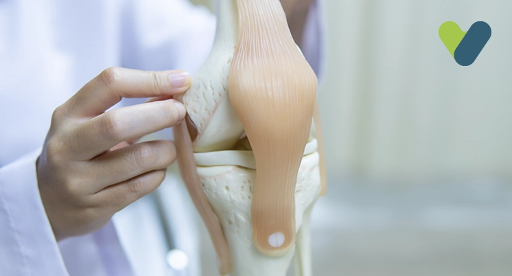
Knee osteotomy
This surgery is usually recommended to patients who experience pain on one side of the knee or have one part of the knee damaged. People who are thin and under 60 are more suited for this surgery.
Jaw osteotomy People whose teeth don’t line up properly are recommended an osteotomy of the lower jaw to rectify its positioning.
Arm osteotomy Sometimes the humerus, radius, or ulna bones in the arm can have an outgrowth or angular issues and cause troubles for the person to go about their daily tasks. The doctor usually recommends such patients to get a corrective osteotomy surgery for their hands. The above-mentioned are some of the common examples, but the surgeon can also perform osteotomy of the big toe, chin, and other bones.
Risks of osteotomy The risks and complications associated with an osteotomy differ slightly depending on the type of osteotomy performed. The following are some of the common risks of an osteotomy:
Blood clot After the surgery, the doctor usually prescribes you a blood thinner and other compression equipment to prevent their blood from clotting and causing damage.
Infection Every surgery poses a risk of infection for the patient; to prevent any infections the patient is given antibiotics during and after the surgery (usually intravenously).
Nerve damage Since the surgeon is operating so close to the bone in an open surgery, there is a small likelihood of mistakes resulting in nerve damage.
Healing failure Although rare, some people may not be able to recover from the osteotomy properly as their bones don’t heal. The screws, plates, or other implants can break or come loose if this happens; additional surgery may be required in such cases.
If you have any doubts about anything related to the procedure, you should consult your doctor before going through with the surgery. It is a good idea to inform them about your medical history and any previous surgeries in the past; some people can also have allergic reactions to anaesthesia.
Post-surgery recovery and rehabilitation
Most people can go home a day or two after the surgery. The doctor gives them care instructions for at-home care along with a prescription for pain medicine and other medicines. It is natural to feel pain after the surgery, but with time, the pain should ideally decrease dramatically as the body heals. Doctors usually prescribe opioids and non-steroidal anti-inflammatory drugs (NSAIDs) to patients.
Post-surgery If the osteotomy was performed on the leg, the patient will require additional support to walk; other parts of the body will usually be stabilised. For example, a brace or cast may be used if the osteotomy surgery was performed on the patient's arm or knee. These measures ensure the patient gradually increases the weight-bearing ability of the new bone. Appropriate guidelines according to your health and recovery will be shared to help you with a proper recovery.
During your recovery and rehabilitation, you will be required to visit the doctor’s clinic periodically. This way, the doctor can monitor your progress closely and ensure proper healing. During these sessions, you may be informed about at-home exercises or be recommended a physiotherapy clinic.
The total recovery time after a hip or femur osteotomy can range anywhere from 3–6 months, during which you will not be allowed to walk initially. Similarly, after a jaw osteotomy, the doctor will likely put you on an all-liquid diet for the first 6 weeks.
The recovery period also depends on the rehabilitation program prescribed by your doctor or physiotherapist. People who ignore their exercise regime can cause damage to their healing bones, which leaves lasting impressions. It is important to strengthen the joint or bone by training the surrounding muscles for stability and strength. In the worst case, you may need additional surgery to rectify the damage if the implants come loose or break.
Outlook for patients Corrective osteotomy surgery is helpful in delaying the progression of arthritis and increasing the longevity of the joint. Young people who have osteotomy surgery instead of a joint replacement can enjoy physical activities after completely healing that otherwise would be out of the question. Corrective osteotomies result in significant pain reduction and increased mobility in patients.
Most patients with osteotomies due to arthritis usually require a joint replacement later in life, but joint replacement can be delayed for several years with the help of corrective osteotomies. If you have any complications after the surgery, including unrelated pain or swelling near the scar and a high fever, you should visit the doctor. You should also pay attention to other factors, such as pain, swelling, mobility, weight-bearing capacity, etc., during the recovery procedure and discuss any concerns with your doctor on your regular visit.


