
A macular hole is seen within the retina of the eye when the macula’s nerve cells get de-linked and start pulling away from the eye’s back surface, thereby hindering overall vision. However, such a macular hole in the eye may be treated successfully with the right care and treatments.
Learning More About the Macular Hole
The hole in retina or macular hole in eye happens due to a particular development. The front portion of the eye is home to a lens which helps in focusing images onto the inside of the back of your eye. This zone, known as the retina, is covered with nerve cells which respond to light just in the manner of film in any camera.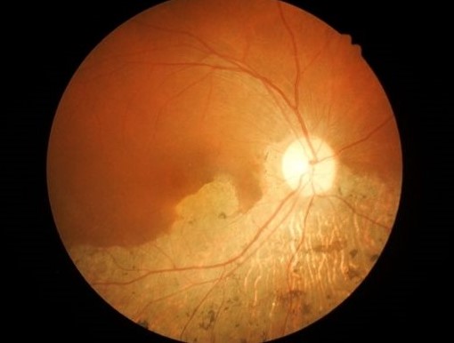
These nerve cells are positioned closely in the middle of the retina where the eye focuses the images that we view and understand. The smaller portion of the retina is known as the macula. At times, the nerve cells in the macula get separated and then pull away from the eye’s back surface, thereby creating a hole. This is known as the macular hole which impairs vision in several ways.
Macular Hole Symptoms and Reasons
Macular holes form due to injuries or specific medical ailments at times, which directly affect the eyes. Nearsightedness is one such reason and it happens owing to traction on the vision center with aging. Some of the other symptoms include the following:- Lower ability to view finer details when an individual is directly viewing any object, irrespective of its distance from the eyes.
- Vision changes which bring a feeling of looking through a thick, foggy or wavy glass or lens.
- Any dark spot which appears in the middle of the vision field.
Testing and Diagnosis
A macular hole is diagnosed through a special procedure where the doctor will use exclusive imaging tests known as optical coherence tomography for getting a cross-sectional insight into the retina. This helps in diagnosis of the macular hole while helping in differentiation of the same from any other similar diagnosis as well.Treatment and Management Aspects
Doctors may not always recommend treatment for macular holes if they are smaller in size and do not cause any major problems with vision. If the eye is otherwise unaffected and fine, then they prefer to wait it out and see. They may recommend frequent eye examinations in these cases in order to ensure that the macular hole does not get bigger or lead to other issues as a result. Patients should maintain their appointments in order to keep track of the situation.Bubble Gas Injection
If there is any reduction in vision and the macular hole is still small, then the doctor may recommend the usage of specific medication or even the injection of a gas bubble into the eye. This will help in traction release and enable the hole to close up in some scenarios. This injection is a non-painful one since the eye is extensively numbed prior to the injection being done. This treatment is only done upon the specific advice of the doctor.Vitrectomy Surgery
If vision gets reduced and the macular hole is not suitable for any such injection, then surgery may also be recommended by the doctor. The surgical method is done with local anesthesia and the patient will stay awake throughout the procedure without feeling anything. The first step is removal of the fluid (gel-like) of the eye or vitreous. This process is called Vitrectomy. The surgeon will create smaller incisions in the eyes for insertion of instruments for removal of the vitreous. Then smaller membranes or tissue fragments/traction near the macular hole may also be removed by the surgeon through forceps.This helps in combating any pulling on the macula that is keeping the hole from closing by itself. Thereafter, the eye fluid is swapped for a sterile gas which helps in keeping the macular hole under the right pressure till it fully heals. Patients will have to ensure a face-downwards position for 1-7 days for keeping this gas bubble firmly in position and closing the hole in question. Surgery is usually successful for 95% of patients with regard to closing the macular hole. Yet, the success rate may come down to about 80% if the right position is not maintained by the patient. The vision regained will vary depending on various aspects and you should consult your doctor about the quantum of visual progress that you may witness.