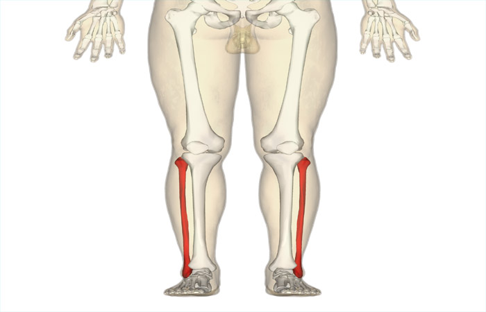
The femur bone is not only the strongest but also the longest bone in the human body. A closer look at femur anatomy will tell you how vital its functions are. Femur bone helps human beings maintain balance, enabling them to move and stand. While femur bone functions are crucial, you should know that serious injuries and accidents lead to breakage of the same. However, another reason behind breakage could be osteoporosis as well. Fracture risks also go up exponentially as a result.
Overview of Femur Bone
You should carefully understand femur anatomy at the outset along with femur bone functions. By the femur, we indicate the thigh bone, i.e., the strongest and longest human bone as mentioned. It has an indispensable role performed in walking, standing and also movement along with body balance. The femur also offers support for crucial muscles, ligaments, tendons and circulatory system components. Since it is the strongest such bone, it can usually absorb the impact of any accident or fall in most cases. However, any such serious accident may lead to fractures/breakage. Surgery is usually needed for bone repair in this case along with ongoing physical therapy to revive overall movement capabilities and strength alike. The femur is also susceptible towards osteoporosis at the same time.Functioning of the Femur Bone
The femur has a job to do on various counts. These include the following:- Holding body weight when you move or stand.
- Connect the ligaments, tendons and muscles in the knees and hips to the rest of the body.
- Stabilizing the body while it moves.
Anatomical Aspects
The femur is the sole bone in the thigh, covering the entire distance between the knee and hip. It has two ends which are rounded and a long middle shaft. This is the classic bone shape that we are familiar with in popular culture. The entire bone resembles a cylinder which has two rounded bumps at both ends of the spectrum. Although it is a single and long bone, the femur bone is made of various components including the following-Femur Proximal Aspect
- The upper femur end links to the hip joint. The proximal end has the neck, head, lesser/greater trochanter and intertrochanteric crest and line.
Femur Shaft
This is the femur portion supporting body weight and forming the thigh structure angling towards the center. The femur shaft contains the gluteal tuberosity, linea aspera, popliteal fossa and pectineal line.Femur Distal Aspect
- The distal or lower femur end comprises the upper portion of the knee joint. It also connects to the patella and tibia. This includes lateral and medial epicondyles along with the lateral and medial condyles and intercondylar fossa.
Femur Bone Disorders and Conditions
The commonest issues impacting the femurs include the following:- Fractures: Bone fractures are only possible in case of serious mishaps and accidents along with massive falls or trauma. Symptoms include swelling, pain, tenderness, discoloration/bruising, inability to move the legs, deformities and bumps which were not seen earlier on the body.
- Osteoporosis: This leads to bones getting weakened, making them highly vulnerable towards fractures that are completely unexpected. Most people do not know about this condition till it leads to bones breaking down. There may not be any visible symptoms. Women and adults who are higher than the age of 50 have higher risks of osteoporosis. You should always consult your doctor in case you suspect the condition.
- Patellofemoral Pain Syndrome (PFPS): This indicates pain hovering near and below the kneecap or patella. This is sometimes known as jumper’s knee or runner’s knee. PFPS may happen due to a lot of things. Symptoms may include pain while squatting, bending down, climbing stairs, sitting with bent knees, popping/crackling sounds in the knees during physical activity, and pain which goes up with changes in workout equipment, intensity of activities and sporting surfaces.
Femur Bone Testing, Treatment and Care
The commonest tests include bone density tests or DXA/DEXA scans. The test helps in measuring the strength of the bones with lower X-ray levels. This is a means of measuring bone density losses with ageing. Femoral fractures may necessitate MRIs, X-rays or CT scans. Treatment options include immobilization (cast/splint) for fractures or surgery for realignment. Osteoporosis treatments will comprise mineral or vitamin supplement consumption, exercising and the right medication.Caring for your femur will mean taking some lifestyle steps that will serve you well on many other counts as well. You should always follow a disciplined lifestyle with a balanced and healthy dietary regime alongside. You should maintain a workout/exercise plan, while consulting your doctor for checkups periodically. This will help you maintain bone health consistently, especially if you are above the age of 50. If you are already past this age group and have a family history of conditions like osteoporosis, make sure you get a bone density scan done.
You should also avoid taking off your seatbelt in the car, while ensuring that you wear safety equipment for all your sporting activities. Ensure that you do not fall or trip over things at home or the office. Do not get up on tables or chairs to reach for any item. Follow the right exercise and diet plan in this regard as well. Use a walker/cane for walking smoothly without any risks of falling down.


