Aarti is a mid-aged housewife living with her beautiful family. She is in her 4th month of pregnancy. Aarti loves doing household work. All she wants to do all day is household chores, cooking, and cleaning for his family. She finds peace in performing household duties. Even after telling her multiple times that doing so much physical work can be strenuous on the baby and negatively affect her pregnancy, she won’t listen to anybody. One day while preparing lunch for her family, Aarti started to get a weird feeling in her stomach. It wasn’t precisely pain, but whatever she felt was new to her. She was immediately taken to her gynaecologist, who prescribed her usg pelvis sonography. She was clueless about what it was as this term was very new to her.
Are you also hearing about it for the first time? Read this blog to know everything about usg pelvis sonography.
What is USG?
When expanded, it is called the ultrasound sonography test, which is a non-invasive diagnostic procedure. It is done to scan the internal organs. High-frequency ultrasound waves are used in it.What Is the USG Pelvis?
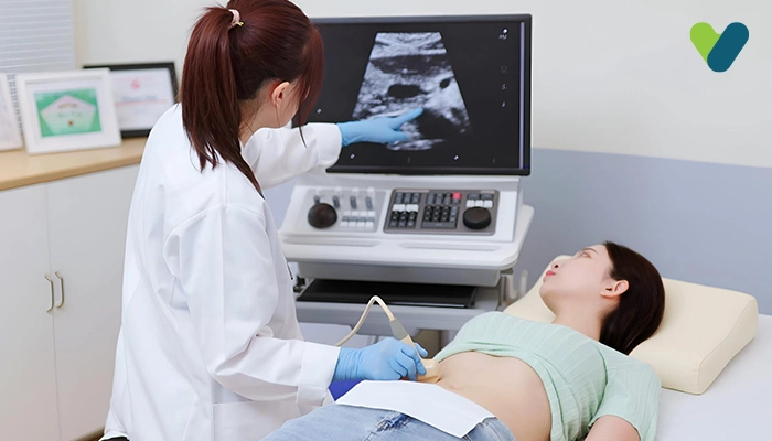
Usg pelvis sonography is usually done to detect any disease in the pelvis or to check a baby's health in the womb of a pregnant woman. The usg pelvis sonography is done to procure images of the pelvis area. It is a non-invasive diagnostic test. It is done to see the pelvis health. It is done to procure real-time images of the pelvis area and to see the movements of the organs inside. It is also done to see the blood flow in the blood vessels.It is done using a transducer that sends out high-frequency ultrasound waves that the human ears can’t hear. The transducer is placed on the skin so that the ultrasound waves can travel in the body to the organs. When the sound wave hits the organ, it bounces off like an echo and travels back to the transducer. The transducer processes these reflected waves are processed by the transducer, which is then converted into an image by the computer. This image shows the examined organs and tissues.
A gel is put on the transducer and the skin to move smoothly without much friction. This gel is also used to eliminate air that is present between the skin and the transducer. This helps in the best sound conduction for usg pelvis sonography.
In usg pelvis sonography, there is no use of radiation. Sometimes your doctor might suggest another kind of ultrasound procedure called the Doppler ultrasound technique. It is a specialised ultrasound procedure. It is used to see the movement of blood flow through arteries and veins, which can be helpful to in usg pelvis sonography.
Procedures for USG Pelvis Sonography
There are three ways through which the procedure of usg pelvis sonography is performed, they are:- Transabdominal – This is done to get the images of the pelvis through the abdomen. The transducer is placed on the stomach, and the gel is used to glide it over the skin to procure pictures of the pelvis.
- Transvaginal – This is done to get the pelvis images by inserting a flexible, long, and thin transducer inside the vagina. The transducer is covered in the gel for ease in insertion and a latex sheath. Through the vagina, the internal organs are assessed.
- Transrectal – This is done to get the pelvis images by inserting a transducer inside the rectum. Through the rectum, the internal organs are assessed.
Importance of USG Pelvis Sonography
It is used to examine the critical pelvic organs in both men and women.In a woman, usg pelvis sonography is used to examine the cervix, ovaries, bladder, vagina, fallopian tube, and uterus.
In a man, usg pelvis sonography is used to examine the prostate gland, the bladder, and the seminal glands.