What are Arteries?
The blood vessels that transport oxygen-rich blood from the heart to the body's tissues are known as arteries. Each artery is a muscular tube with three layers of smooth tissue lining it: The intima is the innermost layer, which is lined with endothelium, a smooth tissue. The media is a layer of muscle that allows arteries to handle the heart's high pressures. The adventitia is a connective tissue that connects arteries to surrounding tissues.
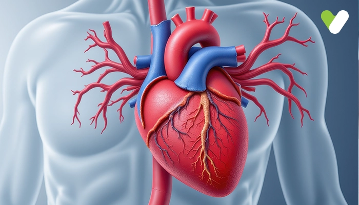
The aorta, which connects the heart's left ventricle to the main high-pressure pipeline, is the biggest artery. The aorta splits into a network of smaller arteries that go all over the body. Arterioles and capillaries are the smallest branches of the arteries. The pulmonary arteries are remarkable in that they transport oxygen-depleted blood from the heart to the lungs under low pressure. The heart muscle receives blood via the coronary arteries. To function, the heart muscle, like all other tissues in the body, requires oxygen-rich blood. Blood that has been drained of oxygen must also be transported. The coronary arteries encircle the heart from the outside in. To supply blood to the heart muscle, little branches delve into the muscle.
2 Main Types of Coronary Arteries
The 2 main coronary arteries are the left main and right coronary arteries:
- Left Main Coronary Artery: Blood is supplied to the left side of the heart muscle by the left main coronary artery (the left ventricle and left atrium). The coronary artery in the left main separates into two branches. The left anterior descending artery is a branch of the left coronary artery that provides blood to the heart's front side. The circumflex artery is a branch of the left coronary artery that surrounds the heart muscle. This artery is responsible for supplying blood to the heart's outer and back sides.
- Right Coronary Artery: The right coronary artery feeds the right ventricle, right atrium, and the sinoatrial and atrioventricular nodes, which control heart rhythm. The right posterior descending artery and the acute marginal artery are two minor branches of the right coronary artery. The right coronary artery, together with the left anterior descending artery, helps to provide blood to the heart's centre or septum.
Physical Structure of Arteries
Smooth muscle, a type of muscle that may be controlled by hormones and particular signals from the nervous system, makes up a large portion of arteries. Collagen fibres make up the artery's outer layer. Smooth muscle and elastic fibres make up the middle layer. The endothelium, or lining, is the innermost layer. The hollow middle of the arteries is where blood travels. Blood pressure might be raised if this hollow-core becomes restricted due to muscle overdevelopment or plaque accumulation. Plaque also reduces the flexibility of the arteries. The tissues that an artery regularly nourishes will die if it ruptures or becomes clogged, as in a stroke or heart attack. Arteries' thick, robust walls allow them to withstand the tremendous pressures found around the heart. Each of the body's major organs has its own set of arteries that are specially designed to provide the necessary supplies. Coronary arteries supply blood to the heart muscle. The aorta branches into the left and right coronary arteries, with the left coronary artery further divided into the circumflex and left anterior descending arteries. During coronary artery bypass graft (CABG) surgery, these four arteries may be replaced. All four arteries are bypassed in a quadruple bypass.
Important Functions of Arteries
Blood is carried away from the heart through arteries. The pulmonary arteries carry oxygen-depleted blood from the right ventricle to the lungs. The left ventricle sends oxygenated blood to the body tissues via systemic arteries. Blood is pumped from the ventricles into huge elastic arteries, which branch into smaller and smaller arteries until tiny arteries known as arterioles are formed. The arterioles are responsible for controlling blood flow into tissue capillaries. At any given time, about 10% of total blood volume is in the systemic arterial system.
Conclusion
Arteries are blood vessels that transport oxygenated blood out from the heart. They have a rough exterior but a smooth inner layer of epithelial cells that permits blood to flow freely. A robust, muscular middle layer exists in arteries, which aids in the pumping of blood throughout the body. They are tube-like in shape and branch out into arterioles to reach organs and tissues. The blood is pushed through the arteries by the heart's pumping contractions.


