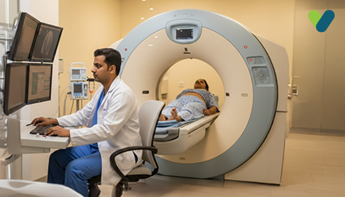
What is an MRI of the dorsal spine?
A diagnostic imaging technique called MRI of the dorsal spine is used to acquire the detailed views of the thoracic or dorsal spine region by employing a powerful magnetic field, radio waves, and a computer. The dorsal spine includes the 12 vertebrae located between the cervical and lumbar regions of the spine.
During this procedure, the individual lies on the table, which goes into the cylindrical scanner. This scanner employs the magnetic field and radio waves to provide detailed images of the spine, which a radiologist or other healthcare professional can view on a computer screen.
This type of MRI can be used to detect a wide range of conditions that affect the dorsal spine, including spinal cord injuries, herniated discs, spinal tumours, infections, and degenerative conditions like spinal stenosis or arthritis. An MRI of the dorsal spine is non-invasive and does not involve radiation, making it a safe and effective tool for diagnosing and monitoring spinal conditions. It is also called a thoracic spine MRI.
Uses of MRI of the Dorsal Spine
The MRI scan of the dorsal spine has multiple applications that enable medical professionals to gain a deeper insight into the spinal issues of their patients. The MRI scan of the dorsal spine can be used to identify various conditions and/ailments, such as:
1. Birth defects of the spinal cord
The MRI of the dorsal spine can detect birth defects of the spinal cord (for example, spina bifida) by producing the detailed images of the spinal cord and surrounding structures. These images can reveal any abnormalities in the size, shape, or alignment of the spinal cord as well as the presence of cysts, tumours, or other growths.
2. Trauma or injury to the dorsal spine
The MRI of the dorsal spine can be used to detect trauma or injury to the dorsal spine. For example, an MRI can reveal herniated discs, fractures, dislocations, or other injuries that may be causing pain or neurological symptoms in the patient.
3. Compression or inflammation of the spinal cord and nerves
The MRI of the dorsal spine can be utilised to identify herniated discs, tumours, cysts, or other growths that may be putting pressure on the spinal cord or nerves. It can also reveal any inflammation or swelling in the spinal cord or surrounding tissues.
4. Spinal cord infections
The MRI of the dorsal spine can reveal inflammation or swelling in the spinal cord or surrounding tissues, which can be a sign of infection. It can also be performed to identify abscesses, which are collections of pus that can form as a result of infection.
5. Spinal cord tumours
Dorsal spine MRI can reveal the presence of tumours or cysts that may be affecting the spinal cord. It can also provide information about the size, location, and characteristics of the tumour, which can help in planning a particular treatment.
6. Joint or disc disease
The MRI of the dorsal spine can be employed to detect the signs of joint damage, such as bone spurs, cartilage degeneration, and inflammation. Disc disorders, such as herniated or bulging discs, can also be detected with a dorsal spine MRI. This MRI can show the size and location of the disc abnormality as well as is any pressure that is exerted on nearby nerves or spinal cord.
Preparation for the MRI of the Dorsal Spine
Before undergoing an MRI of the dorsal spine, some preparation may be required. Here are some general steps that should be followed during this scanning procedure:
- The patient must inform their healthcare provider if they have any metal implants, such as a pacemaker, artificial joints, or metal fragments in your body, as these devices may interfere with the MRI scan.
- The patient must remove any metal objects you may be wearing, such as watches, jewellery, hairpins, or glasses.
- The patient must wear loose, comfortable clothing without any metal fasteners or zippers.
- The patient should avoid eating or drinking for a few hours before the MRI scan if they are going to have contrast material injected, which is a special dye used to enhance the images.
- The patient must let the healthcare provider know if they are pregnant or think they may be pregnant, as MRI scans are generally avoided during the first trimester.
- The patient should fill out any medical history or symptom-related forms or questionnaires.
Any other specific instructions given by the healthcare provider or the imaging centre must be obeyed, as these may vary depending on the type of MRI machine being used and the specific protocol for the scanning procedure.

