A Cardiac PET scan is a diagnostic tool used to evaluate your heart's blood flow. A tiny quantity of a radioactive tracer is put into a vein during this test. There is no colour or contrast in the tracer. The radiation emitted by the tracer is detected by a specialised camera called a PET scanner, which then generates the digital images of your heart. Compared with the, cardiac PET/CT offers better accuracy, less radiation, and enhanced efficiency in diagnosing coronary artery disease (SPECT). Patients with a high body mass index (BMI), excess breast/chest wall tissue, breast implants, or pleural or pericardial effusions may benefit the most from a cardiac PET Scan.
A PET scan is generally accessible. However, a cardiac PET scan is typically not an option for those who are pregnant, nursing, or have recently experienced an acute medical problem. You will have a thorough discussion about this with your ordering doctor.
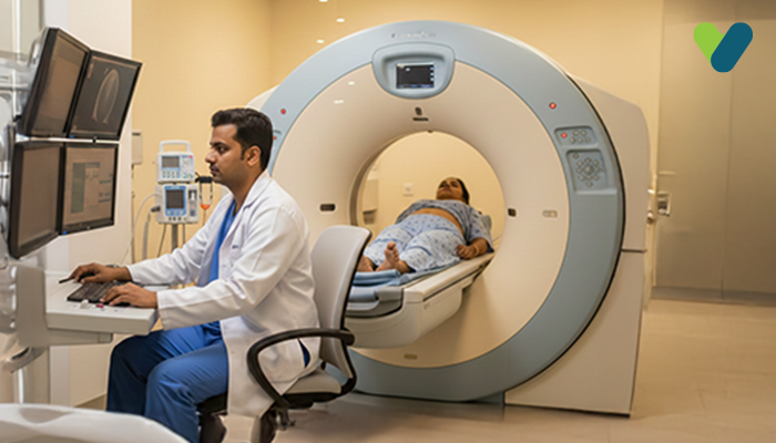
What is Cardiac PET Scan?
A cardiac PET scan is a medical imaging that enables medical practitioners to evaluate how well your heart functions by showing the blood flow to its muscle. During the operation, a tiny quantity of radioactive tracer is put into your bloodstream; it, then, travels to the heart and emits signals that the PET scanner can pick up. Cardiac disorders like coronary artery disease, myocardial infarction, and heart failure are detected by doctors with the help of the images produced during the Cardiac PET CT scan. They also provide detailed information of the metabolic activities of your heart muscle. The non-invasive scanning process can guide physicians in determining your heart conditions and the range of treatment you need.In addition to the test, a drug (vasodilator) is used to dilate your blood vessel. Your doctor can determine if your heart receives enough blood when you are active compared to when you are at rest because of the test and medicine.
When do you need a heart PET scan?
If you are exhibiting heart-related symptoms, your doctor might recommend a heart PET scan. The common symptoms of heart disease include:- Unsteady heartbeat (arrhythmia)
- Chest pain
- Chest tightness
- Difficulty in breathing
- Excessive weakness
- Excessive perspiration
Preparation before Cardiac PET Scan
You will receive thorough instructions from your physician on how to get ready for your heart PET scan. All medicines that you are taking, including over-the-counter medicines and dietary supplements, should be known to the doctor before the scan. You could be requested to fast for up to 8 hours before the surgery, though drinking water might be permitted depending on your medical history and other circumstances. If you are pregnant, suspect pregnancy, or are breastfeeding, you must tell your doctor right away. Your unborn kid may be at risk by this test. Your doctor should provide you with some special instructions for the test if you have a medical condition like diabetes because fasting before the test could impact your blood sugar levels.Procedure of Cardiac PET Scan
You must wear a medical gown and remove all outerwear from the waist up. An EKG or ECG (echocardiogram) will be performed before and during the test to monitor your heart's electrical activity. Adhesive patches called electrodes will cover your body to record the EKG, and your blood pressure will be measured before the test. An IV will be inserted into your arm to administer a tracer and medicine during the test. While lying on the examination table, you will remain still as a camera captures images. A small amount of radioactive tracer will be given to you through your IV to monitor your blood flow while resting.After approximately 20 minutes, the first set of images will be taken. Then, your IV will be infused with a vasodilator drug to simulate exercise, and a second tracer dose will be administered to monitor blood flow while you move around.
Your physician will compare the results of both scans. The medical staff will ask you how you feel throughout the examination, so be sure to let them know if you have any chest, arm, or jaw discomfort, strange symptoms, shortness of breath, a racing heart, or dizziness. Finally, the IV will be removed from your arm after the conclusion of the procedure.
After the Cardiac PET Scan
After the test, you are advised to drink a lot of water to assist your body in flushing the tracers out. All tracers typically leave your body naturally within two days.The images will be interpreted by professionals like doctors with training in inferring PET scans, who will then provide your doctor with the results. The outcomes will then be discussed with you by your doctor during a subsequent visit.
What Conditions Can a Cardiac PET Scan Detect?
Your PET scan images will be interpreted by a professional with training in reading PET scans, who will then provide your doctor with the results. The outcomes will then be discussed with you by your doctor during a subsequent visit.A heart PET scan is a diagnostic test that produces a detailed heart image, allowing doctors to identify the areas of decreased blood flow. It also provides the information of any damage or scar tissue in the heart. When the arteries that feed the heart with blood and oxygen become blocked, constricted, or hardened, it is possible to identify coronary artery disease (CAD) by utilising pictures. There are various treatment options for CAD, which include catheter-assisted angioplasty, which uses a balloon to widen the artery, and coronary bypass surgery, which involves creating a new blood vessel.
Another condition that can be diagnosed using the heart PET scan is heart failure, which can be cause by various factors, such as heart attack, valve disease, or abnormal heart rhythms. Heart failure can be treated by medications, surgery, or medical devices like pacemakers or defibrillators. Your doctor may recommend further testing and treatment based on your results.
Risks of Cardiac PET Scan
Although radioactive tracers are used throughout the scan, your exposure is very low. The American College of Radiology Imaging Network states that the exposure level is too low to significantly impact your body's regular functions.A heart PET scan presents additional risks, such as:
- Muscle aches from laying on the hard test table, which may make you feel uneasy if you have claustrophobia.
- Radiation might be dangerous for an unborn child. Your doctor could suggest an alternative test if you think you might be pregnant or nursing.