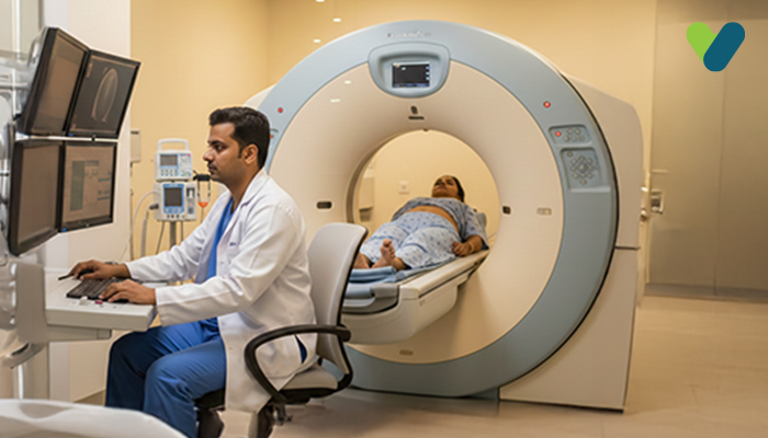
To diagnose various gastrointestinal conditions, doctors often use a specialised radiologic (X-ray) examination called the barium enema, which allows them to visualise the lower intestinal tract (colon, anus, and rectum). This imaging technique involves the administration of barium sulphate solution, followed by X-ray imaging to capture detailed images of the lower gastrointestinal (GI) tract.
Additionally, barium enema is known as:
- Lower gastrointestinal tract radiography
- Colon X-ray
- Lower GI X-ray
- Lower GI exam
The purpose of the Barium Enema Test
The barium enema test is used for the investigation and diagnosis of the underlying reasons for abdominal discomfort. Previously, medical professionals utilised lower GI exam (barium enema) as a diagnostic tool to explore the underlying factors contributing to abdominal symptoms. However, with the advent of more precise imaging tests like CT scans, the prominence of barium enema has diminished.The doctor may recommend a barium enema exam to identify the root cause if you experience symptoms such as rectal bleeding, abdominal pain, bowel habit alterations, chronic diarrhoea, an unknown cause of weight loss, or chronic constipation. Additionally, your doctor might have previously prescribed a barium enema radiology (X-ray) to investigate for conditions such as polyps (abnormal growths) as component of the screening process for colorectal cancer or to detect signs of chronic inflammatory conditions (for example, inflammatory bowel disease).
It is important to note that newer imaging tests, such as CT scans, have emerged as more accurate alternatives to barium enema in the present medical landscape.
Understanding Barium Enema
A barium enema is a diagnostic procedure that uses an X-ray exam to identify changes or abnormalities in the large intestine. It involves the administration of a liquid containing barium, a metallic substance, into the rectum through a small tube. The barium coats the lining of the colon, enhancing the visibility of soft tissues during the X-ray imaging process.To further enhance the image quality, air may be introduced into the colon during the procedure. This air-contrast technique, also called a double-contrast barium enema, expands the colon, resulting in clearer images.
Preparation for a Barium Enema
You will be given specific instructions on how to ensure your colon is empty before undergoing barium enema radiology. This is crucial to avoid any potential obscuring of X-ray pictures or misinterpretation of findings.The preparatory steps may include:
- Following a specialised diet: One day prior to the barium enema exam, you will be advised to adhere to a specific diet. This may involve abstaining from solid foods and consuming only clear fluids such as tea, coffee without cream or milk, water, clear broth, and carbonated beverages that are transparent.
- Fasting after midnight: Typically, you will be instructed to refrain from eating or drinking after midnight on the day of the exam.
- Administering a laxative: You will be provided with a laxative liquid or pill, the night prior to the test. This laxative will facilitate the complete evacuation of your colon.
- Using a cleansing (enema) kit: This kit can be used either the evening prior to the test or a few hours prior to the scheduled procedure. It contains a specialised solution that helps cleanse your colon by eliminating any residual substances.
- Consulting about medications: It is essential to consult your healthcare provider about the medications you regularly take at least one week prior to the exam. Your doctor may recommend discontinuing certain medications before the procedure.
Procedure for Barium Enema X-ray
During the barium enema procedure, you will be provided with a hospital gown and requested to remove accessories and eyewear, except for removable dental bridges or implants. A radiology technologist under the guidance of a radiologist will perform the exam. The steps of the procedure are as follows:- To initiate the exam, you will be asked to lie laterally (on one side) on an examination table. To ensure that your colon is completely empty (cleansed), an initial X-ray image will be captured. Next, an enema tube (well-lubricated) will be gently introduced through your anus, through which a barium solution will be delivered into the colon. In cases where a double contrast technique of barium enema is being performed, the same enema tube will also be used for introducing air into the rectum.
- The enema tube used for barium delivery is equipped with a balloon (small) at its tip. This balloon assists in retaining the barium inside your body by positioning it at the opening of your rectum. As the barium fills your colon, you may experience a bowel evacuation and may also encounter abdominal cramping. It is important to keep the tube in position while taking slow, deep breaths to help relax.
- Throughout the procedure, you may be instructed to change and maintain different postures on the examination table. This ensures that the barium thoroughly coats your colon, thereby allowing the radiologist doctor to examine it from multiple angles. At times, you might be instructed to briefly pause your breath.
- The radiologist doctor may apply gentle pressure to your abdominal and pelvic region, manoeuvring your colon to enhance the visualization on a screen connected to the X-ray scanner. Multiple X-ray images of the colon will be captured from different angles.
- On average, a barium enema test takes somewhere between 30 and 60 minutes to complete.
After the Barium Enema
Following the completion of the exam, the barium from the colon will be primarily expelled via the enema tube. Once the tube is taken out, you can use the restroom to eliminate any remaining air and barium. Any abdominal pain or discomfort experienced during the procedure typically subsides quickly, allowing you to resume your regular diet and daily activities immediately.It is common to have white or pale-coloured bowels for a couple of days since your body’s system naturally eliminates any residual barium from the colon. Barium can lead to constipation, so it is recommended to increase your water intake after the scan. Your doctor can suggest using a laxative if necessary.
If you encounter difficulties in passing gas or having a normal bowel movement beyond two days post-exam, or if the bowels (stool) do not return to their usual colour in a couple days, it is recommended to consult your doctor for further evaluation and guidance.
Does a Barium Enema Cause Pain?
It is essential to note that while barium enema is unlikely to be painful, the experience may bring about a sense of uneasiness. Sensations of discomfort may arise as air is gently pumped into the bowel during the test. Some individuals may encounter bloating or even stomach cramps after the procedure.Interpreting the results
After the examination concludes, the radiologist will compile a comprehensive report summarising the findings and promptly send it to your healthcare provider. Your provider will then review the results and discuss any potential need for further investigations or interventions that might be necessary.Negative Result: If the radiologist doctor finds no abnormalities within the colon during the barium enema test, it is considered a negative result. This implies the absence of any issues that were discernible.
Positive Result: Conversely, a positive result denotes that the radiologist has identified some abnormalities or irregularities in the colon during the barium enema test. Based on the nature of these findings, your doctor might advise additional tests, for instance, a colonoscopy, in order to conduct a more comprehensive examination. They may also perform biopsies if necessary or remove any identified abnormalities.
In cases where your doctor has concerns regarding the clarity or reliability of the obtained X-ray pictures, they may advise repeating the barium enema procedure or suggest alternative diagnostic tests to ensure accurate and dependable results.
Risks of barium enema radiology
It is crucial to be aware of the potential risks associated with barium enema radiology. However, the benefits of obtaining an accurate diagnosis outweigh the risks associated with the minimal amount of radiation exposure during the test.Pregnancy is a sensitive time, and the utmost care should be taken when considering the use of X-rays. It is crucial to understand that these medical procedures are generally not advised for expectant mothers due to the potential risks linked to radiation exposure, which may have adverse effects on the growing foetus.
In cases where there is a possibility of a tear or perforation in the colon, your doctor may choose a contrast solution containing iodine. This alternative solution carries fewer risks of complications if it happens to leak from the colon.
The barium solution can sometimes trigger an immune response that can result in allergies. It is important to inform your healthcare provider about any allergies to ensure appropriate measures are taken.
Other uncommon complications that can arise from a barium enema include:
- Inflammation of the tissues surrounding the colon
- Obstruction of the gastrointestinal tract
- Perforation of the colon
Recovery and Follow-up
After a barium enema, patients are typically advised to rest as they may experience temporary discomfort or bloating. It is crucial to follow any post-procedure care instructions provided by the healthcare team, including drinking plenty of fluids to flush out the contrast medium. In some cases, additional examinations or follow-up appointments may be recommended to further evaluate any detected abnormalities or monitor the progress of a known condition.Alternatives to Barium Enema
A notable substitute for barium enema is colonoscopy, a method that involves the insertion of a flexible tube equipped with a camera through the rectum to obtain a comprehensive visual representation of the entire colon.Virtual colonoscopy is another option, which employs advanced imaging techniques such as CT scans to produce a three-dimensional image of the colon.
Each alternative has its own benefits and limitations. The selection of the most suitable procedure relies on diverse factors such as the patient's preferences, medical history, and particular clinical circumstances.

