What is vasa previa?
The term “vasa” comes from Latin, meaning “vessels”, the term “pre” meaning before, and the term “via” meaning way. In simpler words, the vessels that lie before the fetus. These vessels lie in the birth canal, “in the way” of the fetus’s exit.
Vasa previa is a serious pregnancy complication where fetal blood vessels cross or run very close to the internal opening of the cervix. These vessels are unprotected by the umbilical cord or placenta, making them vulnerable to rupture when labor begins or when the membranes break. It is a condition that can lead to severe fetal blood loss and even stillbirth if not diagnosed early. Fortunately, with timely detection and proper vasa previa management, most pregnancies can have positive outcomes.
What are the types of vasa previa?
There are two Vasa Previa types:
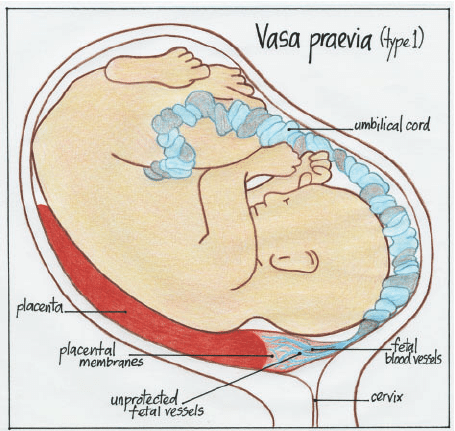
- Type 1 vasa previa: This type occurs when the umbilical cord attaches to the membranes instead of the placenta. This leaves the vessels exposed and unprotected.
- Type 2 vasa previa: This type occurs when the vessels connecting the placenta and a smaller accessory lobe pass over the cervix. In this type, the placenta can have two lobes, too.
- Type 3: This is a less common type, identified recently. In this type, the fetal vessels branch out from the placental edge, cross the membranes, and then return to the placental edge. This type can occur even with a normally inserted cord and without placental abnormalities.
Now, all types are risky. Early vasa previa diagnosis allows doctors to take preventive steps for a safe delivery. The knowledge of these classifications can help you with accurate vasa previa management and narrow down your vasa previa treatment plan.

How serious is vasa previa?
Vasa previa is a very serious obstetric condition, considered high-risk in the medical field. There is a high risk that if the condition goes undetected, it can cause serious harm to the fetus. It can cause rapid fetal blood loss during labor. The severity of this condition lies in the fact that this fetal blood loss takes minutes if the exposed vessels rupture.
This is why early intervention and vasa previa diagnosis are essential. It can help you receive the vasa previa treatment you need and manage your symptoms, drastically improving outcomes.
Modern vasa previa management strategies suggest an early vasa previa diagnosis and planned delivery before labor begins. Today’s prenatal screening processes and improved awareness make it so that the survival rates in diagnosed cases are over 95%. The key is vigilance, accurate diagnosis, and timely vasa previa treatment.
Who does vasa previa affect?
Vasa previa can affect anyone, but some people are at a higher risk than others. The most common and significant vasa previa risk factors include:
- Those who have been pregnant multiple times
- Those who underwent IVF treatment
- Those with placenta previa
- Those experiencing velamentous cord insertion
- Presence of a two-lobed placenta.
Women with these vasa previa risk factors should be closely monitored during pregnancy. The second and third trimesters are the most crucial time. Early ultrasound can detect the condition before complications arise. When you understand these vasa previa risk factors, you can be empowered as an expectant mother to seek proactive care. You can also help your doctor create safer and more effective birth plans through appropriate vasa previa management.
How common is vasa previa?
Vasa previa occurs in approximately 1 in 2500 pregnant women. It is still relatively rare, but its impact can be severe, especially if left undiagnosed and untreated. In India, the exact data is limited because many cases go unreported, and there are differences in access to proper diagnostic equipment across the country. The experts estimate that vasa previa may affect around 1 in 3000 pregnancies in India.
Technology is always advancing. There is increased use of advanced prenatal imaging techniques, like the transvaginal color Doppler ultrasound. These tests have led to more frequent detections of vasa previa in recent years. Awareness campaigns and improved vasa previa management have led to better survival rates for the mother and the child. When you understand how the vasa previa diagnosis works, it can make a significant difference in how you are treated. This knowledge is especially useful in countries where access to reproductive healthcare is still developing.
What are the signs and symptoms of vasa previa?
Vasa previa often doesn’t show symptoms until labor or if the membrane ruptures. As a result, it is often detected during a routine ultrasound, rather than by symptoms. However, in those that do show symptoms, the following symptoms are noticed:
- Painless vaginal bleeding: Typically occurs during late pregnancy
- Abnormal fetal heart rate: This symptom especially develops if the membranes rupture
- Bleeding with a change in fetal heart rate
Vasa previa symptoms can resemble other conditions, like placenta previa. An accurate vasa previa diagnosis is required in this case to rule out other conditions. Recognizing early signs and adhering to your routine ultrasounds can ensure timely vasa previa treatment. Timely treatment also prevents complications, especially life-threatening ones.
What causes vasa previa?
There are several causes of vasa previa. Let’s take a closer look at them:
- Velamentous Cord Insertion: This happens when the umbilical cord attaches to the membranes instead of the placenta, like it’s supposed to. This leaves the fetus’s blood vessels exposed and unprotected. Typically, a gel-like substance called Wharton’s Jelly provides a soft cushion for these blood vessels. A velamentous cord insertion also means that the umbilical cord cannot reach the nutrients from the placenta in a timely manner, making it slow in carrying back the nutrients to the baby.
- Bilobed Placenta: In some vasa previa cases, the placenta splits into two equal-sized lobes. The umbilical cord becomes implanted in either lobe, the membranes, or between both placental lobes. When the lobes split, the blood vessels that connect them lie unprotected and vulnerable over the cervix. This increases the risk of the vessels rupturing during labor. The fetus can start bleeding severely if this happens.
- Succenturiate placenta: This is similar to the bilobed placenta condition, except the two halves are of different sizes. One is significantly smaller than the main placenta. Succenturiate placenta is a rare occurrence, but it has been linked as part of vasa previa causes.
Vasa previa causes also include other health conditions like placenta previa, where the placenta attaches lower in the uterus, covering the cervical opening. Understanding vasa previa causes help in early risk identification. Doctors can apply specific vasa previa management techniques, such as close monitoring and scheduling an early delivery. This helps avoid vessel rupture. Since many of the vasa previa causes are structural and cannot be prevented entirely, awareness and regular screening are your best defenses.
How is vasa previa diagnosed?
Vasa previa diagnosis generally happens during a routine ultrasound, using a transvaginal scan. This specific test allows doctors to see if fetal vessels are crossing over the cervix. The test takes place around 18 to 26 weeks. A transvaginal ultrasound is especially helpful for those women who possess ultrasound risk factors for vasa previa. The color Doppler feature of an ultrasound shows a clearer image of the blood vessels.
If your doctor suspects that you may have vasa previa, you will be called for follow-up scans to confirm the vasa previa diagnosis. An accurate vasa previa diagnosis will help you get the precise vasa previa treatment. Typically, the treatment involves early delivery through C-section. In many places, vasa previa management now includes targeted ultrasounds for patients who are exhibiting vasa previa risk factors.
Your doctor will note the following points:
- Where the placenta is positioned
- Whether the placenta is whole or has multiple lobes
- Where the umbilical cord has attached
What is the treatment for vasa previa?
We saw the vasa previa diagnosis; now, let’s take a look at vasa previa treatment options.
- First, your doctor will schedule an early C-section and closely monitor your condition
- Your doctor may conduct non-stress tests twice a week. These tests will help assess the fetus’s heart rate. These tests are completely safe for both the mother and the baby.
- Corticosteroid medications may be prescribed as part of your vasa previa treatment plan. These medications can help develop the fetus’s lungs in preparation for the C-section
- In the days leading up to your delivery, your doctor may recommend getting yourself admitted for close monitoring. Your doctor will check your medical history, your likelihood of going into labor, and how far away you live from the hospital to decide the exact date
- Your doctor will schedule a C-section at about 34 to 37 weeks. Before this, your doctor will discuss with you the potential risks and benefits of the delivery timing. This is to reduce the risk of complications. Many fetuses born via C-section in early delivery require neonatal care.
How can I reduce my risk of vasa previa?
Since many vasa previa causes are structural and cannot be fully prevented, reducing risk largely depends on early detection. Here are some steps you can take:
- Attend all prenatal visits and schedule regular ultrasounds.
- Discuss any history of placenta previa or abnormal cord attachment with your doctor.
- If you have vasa previa risk factors, such as IVF conception or multiple pregnancy, ask for a targeted Doppler ultrasound.
- Follow your healthcare provider’s advice about vasa previa management and timing of delivery.
While prevention isn’t always possible, being proactive greatly lowers the risk of complications and improves outcomes through effective vasa previa treatment planning.
When should I see my healthcare provider?
You should contact your doctor right away if you experience vaginal bleeding, especially in the second or third trimester, or notice any unusual symptoms like decreased fetal movement. Even if bleeding stops, it’s important to get evaluated. Early vasa previa diagnosis can make all the difference between a high-risk and a healthy pregnancy outcome.
If you fall into a category with known vasa previa risk factors, your provider may recommend extra scans and monitoring. Discussing your vasa previa management plan ensures that both you and your baby stay safe through timely intervention and personalized vasa previa treatment.
Connect with a gynaecologist online for timely vasa previa diagnosis and personalized pregnancy care to keep you and your baby healthy.
Key Take aways
- Vasa previa definition in OBG: It refers to a condition where fetal blood vessels cross over the cervix, unprotected.
- Vasa previa types: There are two main vasa previa types, both requiring careful prenatal screening and planning. Recently, a third type has been found, but it is a rare occurrence.
- Vasa previa diagnosis: Early vasa previa diagnosis through ultrasound is key to preventing severe complications.
- Vasa previa and placenta previa: These are distinct conditions, though both can cause bleeding during pregnancy.
- Vasa previa risk factors: Include multiple pregnancies, IVF conception, Placenta previa, etc. Seeking prompt care improves outcomes.
- Vasa previa management: Effective vasa previa management includes hospitalization, monitoring, and planned cesarean delivery.
- Vasa previa treatment: With timely vasa previa treatment, survival rates for babies now exceed 95%.